Magnesium »
PDB 3gx3-3hd1 »
3gxz »
Magnesium in PDB 3gxz: Crystal Structure of Cyanovirin-N Complexed to Oligomannose-9 (Man-9)
Protein crystallography data
The structure of Crystal Structure of Cyanovirin-N Complexed to Oligomannose-9 (Man-9), PDB code: 3gxz
was solved by
I.Botos,
B.R.O'keefe,
S.R.Shenoy,
P.H.Seeberger,
M.R.Boyd,
A.Wlodawer,
with X-Ray Crystallography technique. A brief refinement statistics is given in the table below:
| Resolution Low / High (Å) | 28.40 / 2.50 |
| Space group | P 41 21 2 |
| Cell size a, b, c (Å), α, β, γ (°) | 61.512, 61.512, 147.950, 90.00, 90.00, 90.00 |
| R / Rfree (%) | 24.6 / 32.3 |
Magnesium Binding Sites:
The binding sites of Magnesium atom in the Crystal Structure of Cyanovirin-N Complexed to Oligomannose-9 (Man-9)
(pdb code 3gxz). This binding sites where shown within
5.0 Angstroms radius around Magnesium atom.
In total only one binding site of Magnesium was determined in the Crystal Structure of Cyanovirin-N Complexed to Oligomannose-9 (Man-9), PDB code: 3gxz:
In total only one binding site of Magnesium was determined in the Crystal Structure of Cyanovirin-N Complexed to Oligomannose-9 (Man-9), PDB code: 3gxz:
Magnesium binding site 1 out of 1 in 3gxz
Go back to
Magnesium binding site 1 out
of 1 in the Crystal Structure of Cyanovirin-N Complexed to Oligomannose-9 (Man-9)
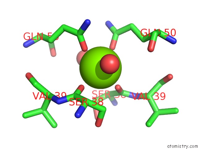
Mono view
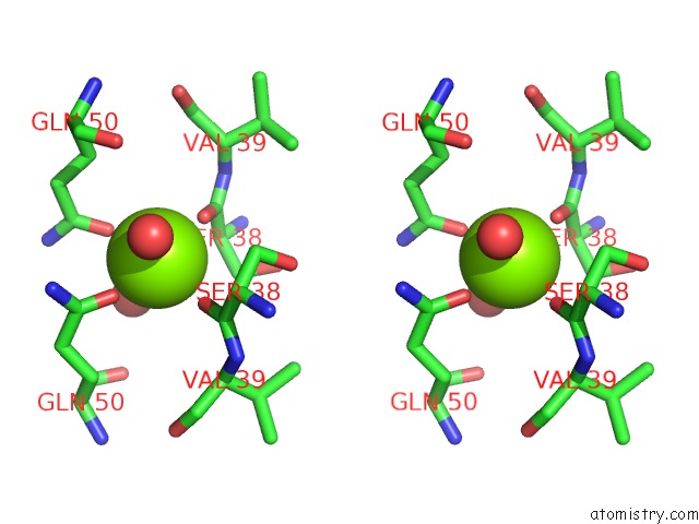
Stereo pair view
Mono view
Stereo pair view
A full contact list of Magnesium with other atoms in the Mg binding
site number 1 of Crystal Structure of Cyanovirin-N Complexed to Oligomannose-9 (Man-9) within 5.0Å range:
|
Reference:
I.Botos,
B.R.O'keefe,
S.R.Shenoy,
L.K.Cartner,
D.M.Ratner,
P.H.Seeberger,
M.R.Boyd,
A.Wlodawer.
Structures of the Complexes of A Potent Anti-Hiv Protein Cyanovirin-N and High Mannose Oligosaccharides J.Biol.Chem. V. 277 34336 2002.
ISSN: ISSN 0021-9258
PubMed: 12110688
DOI: 10.1074/JBC.M205909200
Page generated: Wed Aug 14 15:00:03 2024
ISSN: ISSN 0021-9258
PubMed: 12110688
DOI: 10.1074/JBC.M205909200
Last articles
Zn in 9MJ5Zn in 9HNW
Zn in 9G0L
Zn in 9FNE
Zn in 9DZN
Zn in 9E0I
Zn in 9D32
Zn in 9DAK
Zn in 8ZXC
Zn in 8ZUF