Magnesium »
PDB 3gx3-3hd1 »
3h87 »
Magnesium in PDB 3h87: RV0301 RV0300 Toxin Antitoxin Complex From Mycobacterium Tuberculosis
Protein crystallography data
The structure of RV0301 RV0300 Toxin Antitoxin Complex From Mycobacterium Tuberculosis, PDB code: 3h87
was solved by
A.Min,
M.R.Sawaya,
D.Cascio,
D.Eisenberg,
Integrated Center For Structureand Function Innovation (Isfi),
with X-Ray Crystallography technique. A brief refinement statistics is given in the table below:
| Resolution Low / High (Å) | 60.52 / 1.49 |
| Space group | P 41 21 2 |
| Cell size a, b, c (Å), α, β, γ (°) | 85.557, 85.557, 155.611, 90.00, 90.00, 90.00 |
| R / Rfree (%) | 15.6 / 17.4 |
Magnesium Binding Sites:
The binding sites of Magnesium atom in the RV0301 RV0300 Toxin Antitoxin Complex From Mycobacterium Tuberculosis
(pdb code 3h87). This binding sites where shown within
5.0 Angstroms radius around Magnesium atom.
In total only one binding site of Magnesium was determined in the RV0301 RV0300 Toxin Antitoxin Complex From Mycobacterium Tuberculosis, PDB code: 3h87:
In total only one binding site of Magnesium was determined in the RV0301 RV0300 Toxin Antitoxin Complex From Mycobacterium Tuberculosis, PDB code: 3h87:
Magnesium binding site 1 out of 1 in 3h87
Go back to
Magnesium binding site 1 out
of 1 in the RV0301 RV0300 Toxin Antitoxin Complex From Mycobacterium Tuberculosis
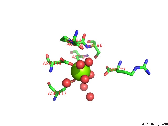
Mono view
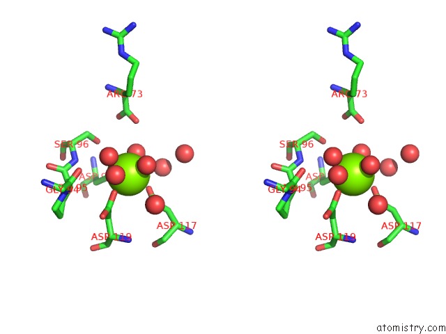
Stereo pair view
Mono view
Stereo pair view
A full contact list of Magnesium with other atoms in the Mg binding
site number 1 of RV0301 RV0300 Toxin Antitoxin Complex From Mycobacterium Tuberculosis within 5.0Å range:
|
Reference:
A.B.Min,
L.Miallau,
M.R.Sawaya,
J.Habel,
D.Cascio,
D.Eisenberg.
The Crystal Structure of the RV0301-RV0300 Vapbc-3 Toxin-Antitoxin Complex From M. Tuberculosis Reveals A Mg(2+) Ion in the Active Site and A Putative Rna-Binding Site. Protein Sci. V. 21 1754 2012.
ISSN: ISSN 0961-8368
PubMed: 23011806
DOI: 10.1002/PRO.2161
Page generated: Wed Aug 14 15:06:02 2024
ISSN: ISSN 0961-8368
PubMed: 23011806
DOI: 10.1002/PRO.2161
Last articles
Zn in 9MJ5Zn in 9HNW
Zn in 9G0L
Zn in 9FNE
Zn in 9DZN
Zn in 9E0I
Zn in 9D32
Zn in 9DAK
Zn in 8ZXC
Zn in 8ZUF