Magnesium »
PDB 3gx3-3hd1 »
3h8a »
Magnesium in PDB 3h8a: Crystal Structure of E. Coli Enolase Bound to Its Cognate Rnase E Recognition Domain
Enzymatic activity of Crystal Structure of E. Coli Enolase Bound to Its Cognate Rnase E Recognition Domain
All present enzymatic activity of Crystal Structure of E. Coli Enolase Bound to Its Cognate Rnase E Recognition Domain:
4.2.1.11;
4.2.1.11;
Protein crystallography data
The structure of Crystal Structure of E. Coli Enolase Bound to Its Cognate Rnase E Recognition Domain, PDB code: 3h8a
was solved by
S.Nurmohamed,
B.F.Luisi,
with X-Ray Crystallography technique. A brief refinement statistics is given in the table below:
| Resolution Low / High (Å) | 24.89 / 1.90 |
| Space group | P 21 21 21 |
| Cell size a, b, c (Å), α, β, γ (°) | 103.883, 110.207, 160.267, 90.00, 90.00, 90.00 |
| R / Rfree (%) | 18 / 22.6 |
Magnesium Binding Sites:
The binding sites of Magnesium atom in the Crystal Structure of E. Coli Enolase Bound to Its Cognate Rnase E Recognition Domain
(pdb code 3h8a). This binding sites where shown within
5.0 Angstroms radius around Magnesium atom.
In total 2 binding sites of Magnesium where determined in the Crystal Structure of E. Coli Enolase Bound to Its Cognate Rnase E Recognition Domain, PDB code: 3h8a:
Jump to Magnesium binding site number: 1; 2;
In total 2 binding sites of Magnesium where determined in the Crystal Structure of E. Coli Enolase Bound to Its Cognate Rnase E Recognition Domain, PDB code: 3h8a:
Jump to Magnesium binding site number: 1; 2;
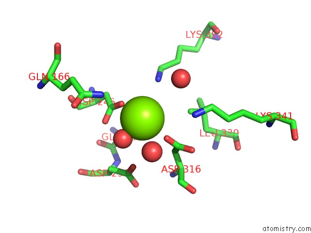
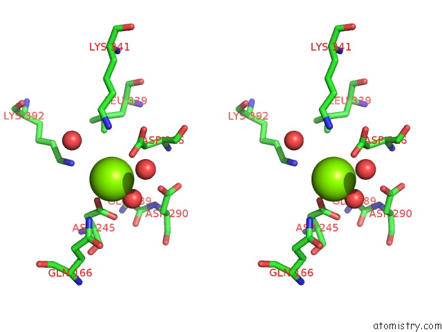
Magnesium binding site 1 out of 2 in 3h8a
Go back to
Magnesium binding site 1 out
of 2 in the Crystal Structure of E. Coli Enolase Bound to Its Cognate Rnase E Recognition Domain

Mono view

Stereo pair view
Mono view
Stereo pair view
A full contact list of Magnesium with other atoms in the Mg binding
site number 1 of Crystal Structure of E. Coli Enolase Bound to Its Cognate Rnase E Recognition Domain within 5.0Å range:
|
Magnesium binding site 2 out of 2 in 3h8a
Go back to
Magnesium binding site 2 out
of 2 in the Crystal Structure of E. Coli Enolase Bound to Its Cognate Rnase E Recognition Domain
Mono view
Stereo pair view
Mono view
Stereo pair view
A full contact list of Magnesium with other atoms in the Mg binding
site number 2 of Crystal Structure of E. Coli Enolase Bound to Its Cognate Rnase E Recognition Domain within 5.0Å range:
|
Reference:
S.Nurmohamed,
A.R.Mckay,
C.V.Robinson,
B.F.Luisi.
Molecular Recognition Between Escherichia Coli Enolase and Ribonuclease E. Acta Crystallogr.,Sect.D V. 66 1036 2010.
ISSN: ISSN 0907-4449
PubMed: 20823555
DOI: 10.1107/S0907444910030015
Page generated: Wed Aug 14 15:06:04 2024
ISSN: ISSN 0907-4449
PubMed: 20823555
DOI: 10.1107/S0907444910030015
Last articles
Zn in 9MJ5Zn in 9HNW
Zn in 9G0L
Zn in 9FNE
Zn in 9DZN
Zn in 9E0I
Zn in 9D32
Zn in 9DAK
Zn in 8ZXC
Zn in 8ZUF