Magnesium »
PDB 3hd2-3hoz »
3hnd »
Magnesium in PDB 3hnd: Crystal Structure of Human Ribonucleotide Reductase 1 Bound to the Effector Ttp and Substrate Gdp
Enzymatic activity of Crystal Structure of Human Ribonucleotide Reductase 1 Bound to the Effector Ttp and Substrate Gdp
All present enzymatic activity of Crystal Structure of Human Ribonucleotide Reductase 1 Bound to the Effector Ttp and Substrate Gdp:
1.17.4.1;
1.17.4.1;
Protein crystallography data
The structure of Crystal Structure of Human Ribonucleotide Reductase 1 Bound to the Effector Ttp and Substrate Gdp, PDB code: 3hnd
was solved by
J.W.Fairman,
S.R.Wijerathna,
H.Xu,
C.G.Dealwis,
with X-Ray Crystallography technique. A brief refinement statistics is given in the table below:
| Resolution Low / High (Å) | 49.94 / 3.21 |
| Space group | P 21 21 21 |
| Cell size a, b, c (Å), α, β, γ (°) | 73.159, 115.834, 221.305, 90.00, 90.00, 90.00 |
| R / Rfree (%) | 18 / 25.4 |
Magnesium Binding Sites:
The binding sites of Magnesium atom in the Crystal Structure of Human Ribonucleotide Reductase 1 Bound to the Effector Ttp and Substrate Gdp
(pdb code 3hnd). This binding sites where shown within
5.0 Angstroms radius around Magnesium atom.
In total 2 binding sites of Magnesium where determined in the Crystal Structure of Human Ribonucleotide Reductase 1 Bound to the Effector Ttp and Substrate Gdp, PDB code: 3hnd:
Jump to Magnesium binding site number: 1; 2;
In total 2 binding sites of Magnesium where determined in the Crystal Structure of Human Ribonucleotide Reductase 1 Bound to the Effector Ttp and Substrate Gdp, PDB code: 3hnd:
Jump to Magnesium binding site number: 1; 2;
Magnesium binding site 1 out of 2 in 3hnd
Go back to
Magnesium binding site 1 out
of 2 in the Crystal Structure of Human Ribonucleotide Reductase 1 Bound to the Effector Ttp and Substrate Gdp
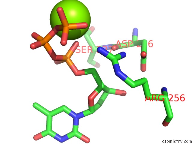
Mono view
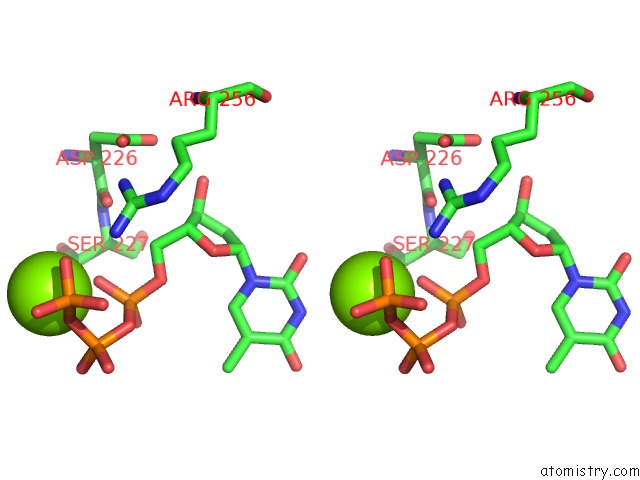
Stereo pair view
Mono view
Stereo pair view
A full contact list of Magnesium with other atoms in the Mg binding
site number 1 of Crystal Structure of Human Ribonucleotide Reductase 1 Bound to the Effector Ttp and Substrate Gdp within 5.0Å range:
|
Magnesium binding site 2 out of 2 in 3hnd
Go back to
Magnesium binding site 2 out
of 2 in the Crystal Structure of Human Ribonucleotide Reductase 1 Bound to the Effector Ttp and Substrate Gdp
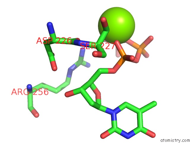
Mono view
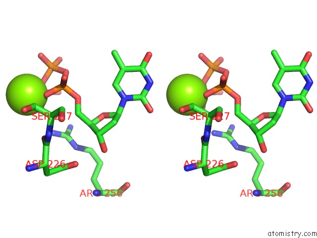
Stereo pair view
Mono view
Stereo pair view
A full contact list of Magnesium with other atoms in the Mg binding
site number 2 of Crystal Structure of Human Ribonucleotide Reductase 1 Bound to the Effector Ttp and Substrate Gdp within 5.0Å range:
|
Reference:
J.W.Fairman,
S.R.Wijerathna,
M.F.Ahmad,
H.Xu,
R.Nakano,
S.Jha,
J.Prendergast,
R.M.Welin,
S.Flodin,
A.Roos,
P.Nordlund,
Z.Li,
T.Walz,
C.G.Dealwis.
Structural Basis For Allosteric Regulation of Human Ribonucleotide Reductase By Nucleotide-Induced Oligomerization. Nat.Struct.Mol.Biol. V. 18 316 2011.
ISSN: ISSN 1545-9993
PubMed: 21336276
DOI: 10.1038/NSMB.2007
Page generated: Wed Aug 14 15:12:43 2024
ISSN: ISSN 1545-9993
PubMed: 21336276
DOI: 10.1038/NSMB.2007
Last articles
Zn in 9MJ5Zn in 9HNW
Zn in 9G0L
Zn in 9FNE
Zn in 9DZN
Zn in 9E0I
Zn in 9D32
Zn in 9DAK
Zn in 8ZXC
Zn in 8ZUF