Magnesium »
PDB 3hd2-3hoz »
3hos »
Magnesium in PDB 3hos: Crystal Structure of the Mariner MOS1 Paired End Complex with Mg
Protein crystallography data
The structure of Crystal Structure of the Mariner MOS1 Paired End Complex with Mg, PDB code: 3hos
was solved by
J.M.Richardson,
M.D.Walkinshaw,
with X-Ray Crystallography technique. A brief refinement statistics is given in the table below:
| Resolution Low / High (Å) | 29.87 / 3.50 |
| Space group | P 1 21 1 |
| Cell size a, b, c (Å), α, β, γ (°) | 121.232, 85.033, 131.273, 90.00, 98.93, 90.00 |
| R / Rfree (%) | 21.9 / 27.9 |
Magnesium Binding Sites:
The binding sites of Magnesium atom in the Crystal Structure of the Mariner MOS1 Paired End Complex with Mg
(pdb code 3hos). This binding sites where shown within
5.0 Angstroms radius around Magnesium atom.
In total only one binding site of Magnesium was determined in the Crystal Structure of the Mariner MOS1 Paired End Complex with Mg, PDB code: 3hos:
In total only one binding site of Magnesium was determined in the Crystal Structure of the Mariner MOS1 Paired End Complex with Mg, PDB code: 3hos:
Magnesium binding site 1 out of 1 in 3hos
Go back to
Magnesium binding site 1 out
of 1 in the Crystal Structure of the Mariner MOS1 Paired End Complex with Mg
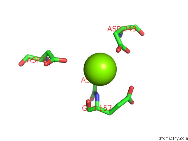
Mono view
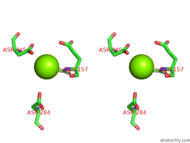
Stereo pair view
Mono view
Stereo pair view
A full contact list of Magnesium with other atoms in the Mg binding
site number 1 of Crystal Structure of the Mariner MOS1 Paired End Complex with Mg within 5.0Å range:
|
Reference:
J.M.Richardson,
S.D.Colloms,
D.J.Finnegan,
M.D.Walkinshaw.
Molecular Architecture of the MOS1 Paired-End Complex: the Structural Basis of Dna Transposition in A Eukaryote Cell(Cambridge,Mass.) V. 138 1096 2009.
ISSN: ISSN 0092-8674
PubMed: 19766564
DOI: 10.1016/J.CELL.2009.07.012
Page generated: Wed Aug 14 15:13:45 2024
ISSN: ISSN 0092-8674
PubMed: 19766564
DOI: 10.1016/J.CELL.2009.07.012
Last articles
Cl in 5YKACl in 5YJS
Cl in 5YJP
Cl in 5YJK
Cl in 5YJM
Cl in 5YHO
Cl in 5YIY
Cl in 5YG4
Cl in 5YHG
Cl in 5YHE