Magnesium »
PDB 3hd2-3hoz »
3hox »
Magnesium in PDB 3hox: Complete Rna Polymerase II Elongation Complex V
Enzymatic activity of Complete Rna Polymerase II Elongation Complex V
All present enzymatic activity of Complete Rna Polymerase II Elongation Complex V:
2.7.7.6;
2.7.7.6;
Protein crystallography data
The structure of Complete Rna Polymerase II Elongation Complex V, PDB code: 3hox
was solved by
J.F.Sydow,
F.Brueckner,
A.C.M.Cheung,
G.E.Damsma,
S.Dengl,
E.Lehmann,
D.Vassylyev,
P.Cramer,
with X-Ray Crystallography technique. A brief refinement statistics is given in the table below:
| Resolution Low / High (Å) | 50.00 / 3.65 |
| Space group | C 2 2 21 |
| Cell size a, b, c (Å), α, β, γ (°) | 221.617, 393.716, 282.576, 90.00, 90.00, 90.00 |
| R / Rfree (%) | 21.2 / 25 |
Other elements in 3hox:
The structure of Complete Rna Polymerase II Elongation Complex V also contains other interesting chemical elements:
| Bromine | (Br) | 1 atom |
| Zinc | (Zn) | 8 atoms |
Magnesium Binding Sites:
The binding sites of Magnesium atom in the Complete Rna Polymerase II Elongation Complex V
(pdb code 3hox). This binding sites where shown within
5.0 Angstroms radius around Magnesium atom.
In total only one binding site of Magnesium was determined in the Complete Rna Polymerase II Elongation Complex V, PDB code: 3hox:
In total only one binding site of Magnesium was determined in the Complete Rna Polymerase II Elongation Complex V, PDB code: 3hox:
Magnesium binding site 1 out of 1 in 3hox
Go back to
Magnesium binding site 1 out
of 1 in the Complete Rna Polymerase II Elongation Complex V
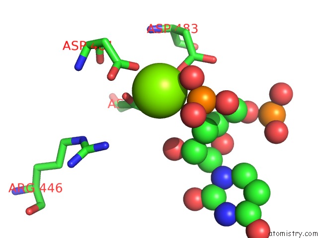
Mono view
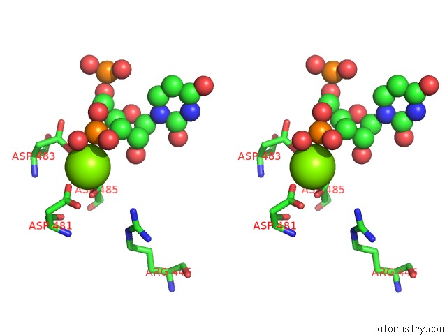
Stereo pair view
Mono view
Stereo pair view
A full contact list of Magnesium with other atoms in the Mg binding
site number 1 of Complete Rna Polymerase II Elongation Complex V within 5.0Å range:
|
Reference:
J.F.Sydow,
F.Brueckner,
A.C.M.Cheung,
G.E.Damsma,
S.Dengl,
E.Lehmann,
D.Vassylyev,
P.Cramer.
Structural Basis of Transcription: Mismatch-Specific Fidelity Mechanisms and Paused Rna Polymerase II with Frayed Rna. Mol.Cell V. 34 710 2009.
ISSN: ISSN 1097-2765
PubMed: 19560423
DOI: 10.1016/J.MOLCEL.2009.06.002
Page generated: Wed Aug 14 15:14:15 2024
ISSN: ISSN 1097-2765
PubMed: 19560423
DOI: 10.1016/J.MOLCEL.2009.06.002
Last articles
Cl in 8BEMCl in 8BGY
Cl in 8BGN
Cl in 8B7U
Cl in 8BFC
Cl in 8BEO
Cl in 8BF4
Cl in 8BDX
Cl in 8BEB
Cl in 8BE9