Magnesium »
PDB 3hp6-3hwx »
3hws »
Magnesium in PDB 3hws: Crystal Structure of Nucleotide-Bound Hexameric Clpx
Protein crystallography data
The structure of Crystal Structure of Nucleotide-Bound Hexameric Clpx, PDB code: 3hws
was solved by
S.E.Glynn,
A.Martin,
T.A.Baker,
R.T.Sauer,
with X-Ray Crystallography technique. A brief refinement statistics is given in the table below:
| Resolution Low / High (Å) | 45.08 / 3.25 |
| Space group | P 21 21 21 |
| Cell size a, b, c (Å), α, β, γ (°) | 54.085, 178.498, 201.369, 90.00, 90.00, 90.00 |
| R / Rfree (%) | 24.3 / 28.2 |
Magnesium Binding Sites:
The binding sites of Magnesium atom in the Crystal Structure of Nucleotide-Bound Hexameric Clpx
(pdb code 3hws). This binding sites where shown within
5.0 Angstroms radius around Magnesium atom.
In total 2 binding sites of Magnesium where determined in the Crystal Structure of Nucleotide-Bound Hexameric Clpx, PDB code: 3hws:
Jump to Magnesium binding site number: 1; 2;
In total 2 binding sites of Magnesium where determined in the Crystal Structure of Nucleotide-Bound Hexameric Clpx, PDB code: 3hws:
Jump to Magnesium binding site number: 1; 2;
Magnesium binding site 1 out of 2 in 3hws
Go back to
Magnesium binding site 1 out
of 2 in the Crystal Structure of Nucleotide-Bound Hexameric Clpx
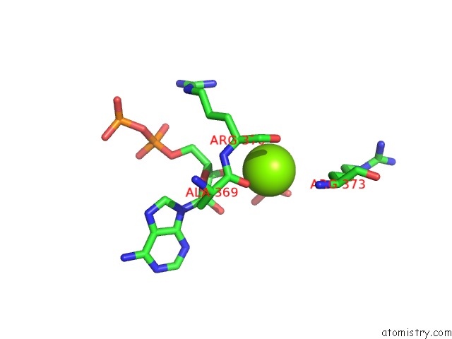
Mono view
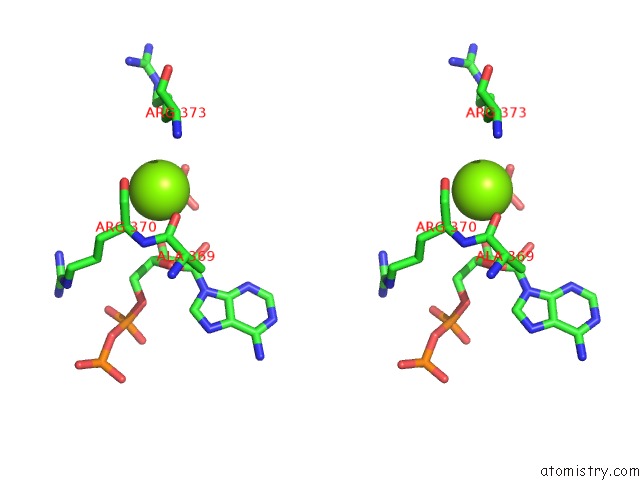
Stereo pair view
Mono view
Stereo pair view
A full contact list of Magnesium with other atoms in the Mg binding
site number 1 of Crystal Structure of Nucleotide-Bound Hexameric Clpx within 5.0Å range:
|
Magnesium binding site 2 out of 2 in 3hws
Go back to
Magnesium binding site 2 out
of 2 in the Crystal Structure of Nucleotide-Bound Hexameric Clpx
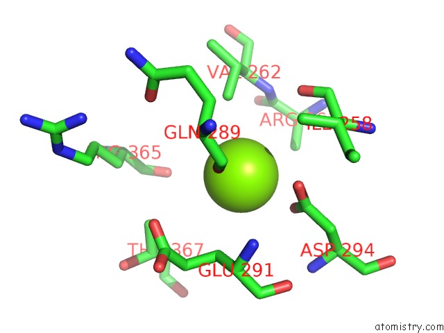
Mono view
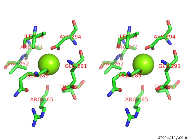
Stereo pair view
Mono view
Stereo pair view
A full contact list of Magnesium with other atoms in the Mg binding
site number 2 of Crystal Structure of Nucleotide-Bound Hexameric Clpx within 5.0Å range:
|
Reference:
S.E.Glynn,
A.Martin,
A.R.Nager,
T.A.Baker,
R.T.Sauer.
Structures of Asymmetric Clpx Hexamers Reveal Nucleotide-Dependent Motions in A Aaa+ Protein-Unfolding Machine. Cell(Cambridge,Mass.) V. 139 744 2009.
ISSN: ISSN 0092-8674
PubMed: 19914167
DOI: 10.1016/J.CELL.2009.09.034
Page generated: Wed Aug 14 15:25:44 2024
ISSN: ISSN 0092-8674
PubMed: 19914167
DOI: 10.1016/J.CELL.2009.09.034
Last articles
Fe in 2YXOFe in 2YRS
Fe in 2YXC
Fe in 2YNM
Fe in 2YVJ
Fe in 2YP1
Fe in 2YU2
Fe in 2YU1
Fe in 2YQB
Fe in 2YOO