Magnesium »
PDB 3hx0-3i5x »
3hyc »
Magnesium in PDB 3hyc: Crystal Structure of E. Coli Phosphatase Yrbi, with Mg, Tetragonal Form
Enzymatic activity of Crystal Structure of E. Coli Phosphatase Yrbi, with Mg, Tetragonal Form
All present enzymatic activity of Crystal Structure of E. Coli Phosphatase Yrbi, with Mg, Tetragonal Form:
3.1.3.45;
3.1.3.45;
Protein crystallography data
The structure of Crystal Structure of E. Coli Phosphatase Yrbi, with Mg, Tetragonal Form, PDB code: 3hyc
was solved by
O.V.Tsodikov,
T.Biswas,
with X-Ray Crystallography technique. A brief refinement statistics is given in the table below:
| Resolution Low / High (Å) | 48.08 / 3.06 |
| Space group | P 41 21 2 |
| Cell size a, b, c (Å), α, β, γ (°) | 122.219, 122.219, 202.173, 90.00, 90.00, 90.00 |
| R / Rfree (%) | 24.2 / 25.7 |
Other elements in 3hyc:
The structure of Crystal Structure of E. Coli Phosphatase Yrbi, with Mg, Tetragonal Form also contains other interesting chemical elements:
| Chlorine | (Cl) | 5 atoms |
Magnesium Binding Sites:
The binding sites of Magnesium atom in the Crystal Structure of E. Coli Phosphatase Yrbi, with Mg, Tetragonal Form
(pdb code 3hyc). This binding sites where shown within
5.0 Angstroms radius around Magnesium atom.
In total 8 binding sites of Magnesium where determined in the Crystal Structure of E. Coli Phosphatase Yrbi, with Mg, Tetragonal Form, PDB code: 3hyc:
Jump to Magnesium binding site number: 1; 2; 3; 4; 5; 6; 7; 8;
In total 8 binding sites of Magnesium where determined in the Crystal Structure of E. Coli Phosphatase Yrbi, with Mg, Tetragonal Form, PDB code: 3hyc:
Jump to Magnesium binding site number: 1; 2; 3; 4; 5; 6; 7; 8;
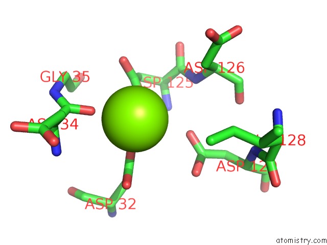
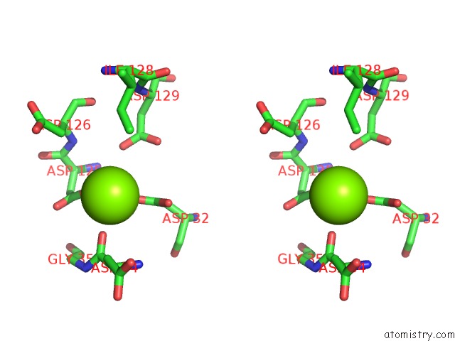
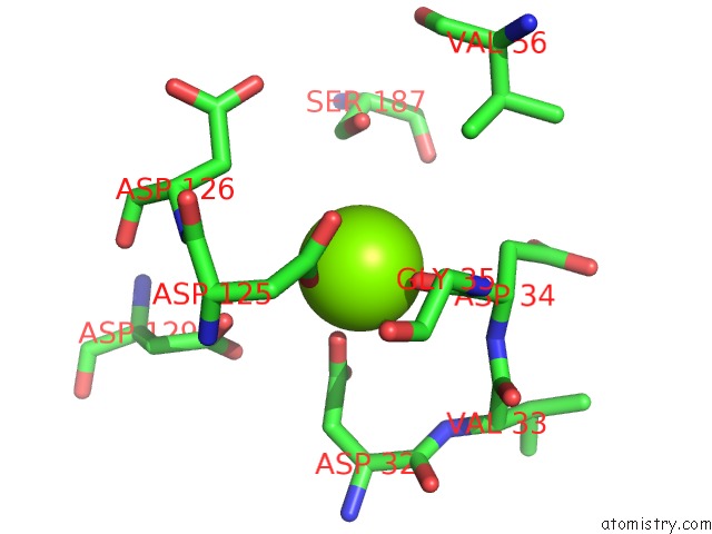
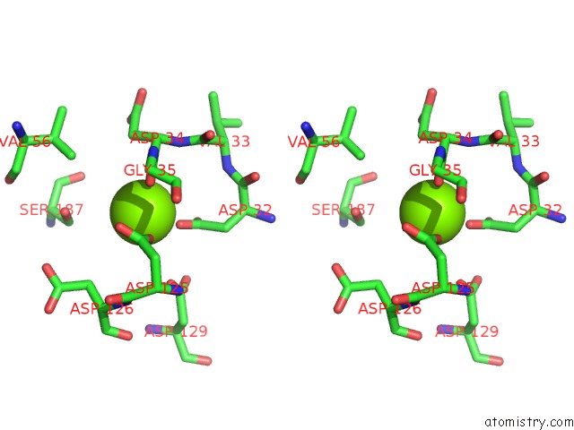
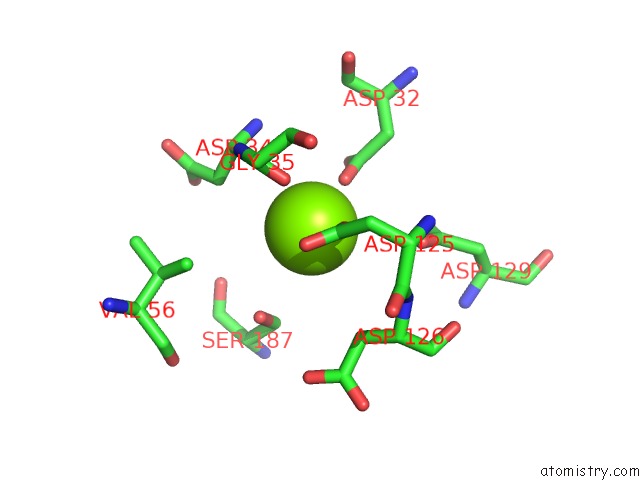
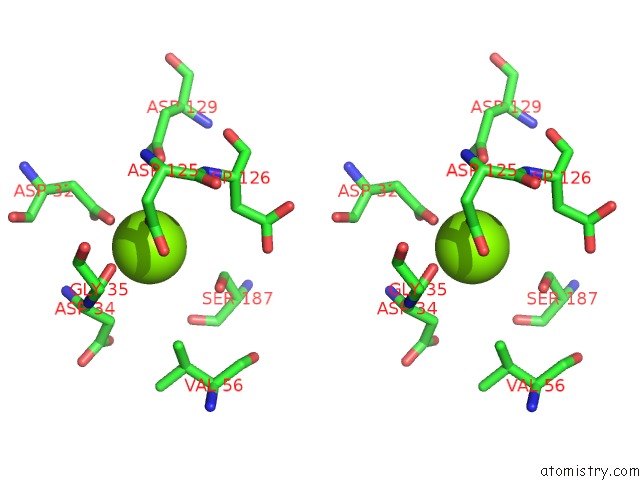
Magnesium binding site 1 out of 8 in 3hyc
Go back to
Magnesium binding site 1 out
of 8 in the Crystal Structure of E. Coli Phosphatase Yrbi, with Mg, Tetragonal Form
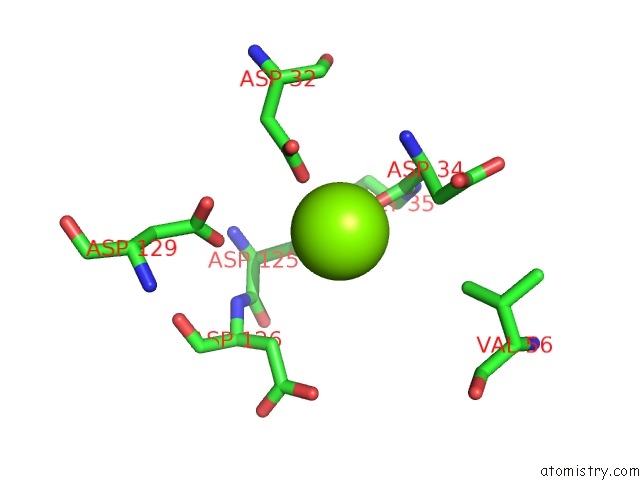
Mono view
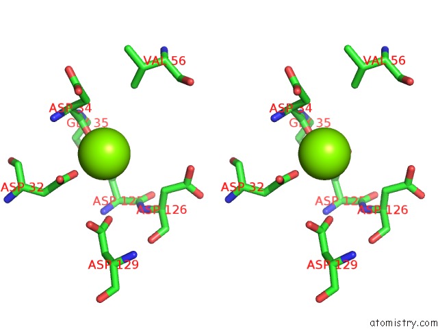
Stereo pair view
Mono view
Stereo pair view
A full contact list of Magnesium with other atoms in the Mg binding
site number 1 of Crystal Structure of E. Coli Phosphatase Yrbi, with Mg, Tetragonal Form within 5.0Å range:
|
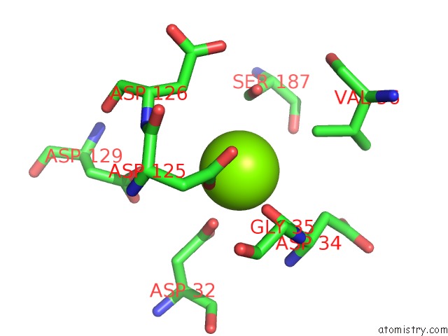
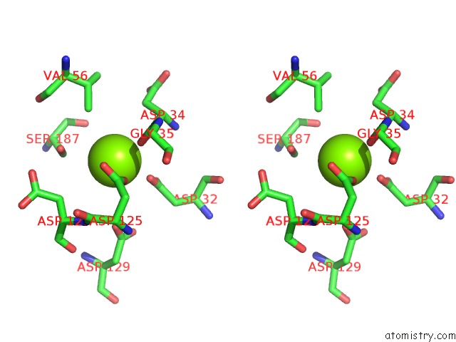
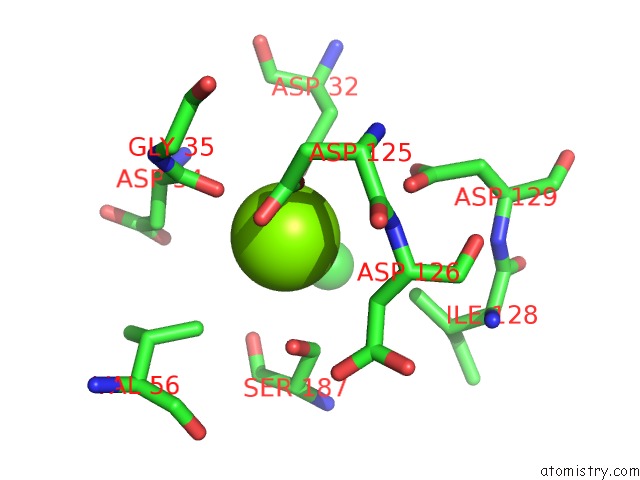
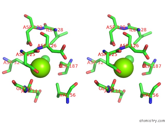
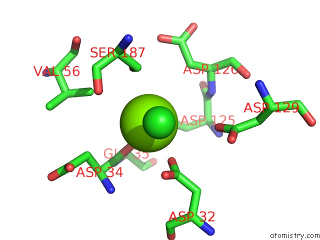
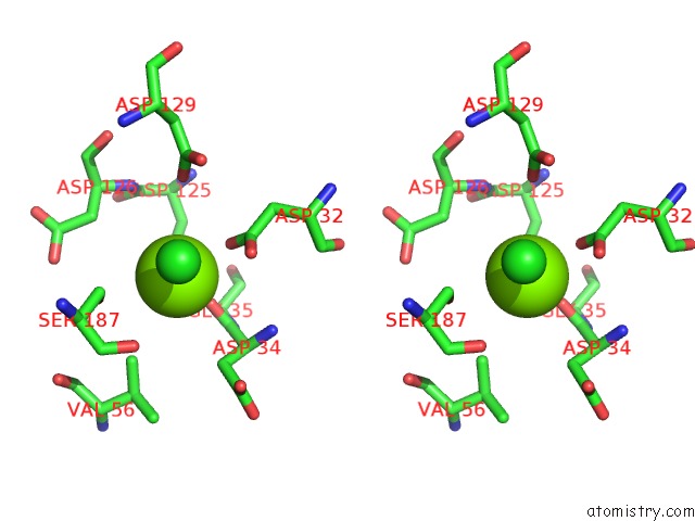
Magnesium binding site 2 out of 8 in 3hyc
Go back to
Magnesium binding site 2 out
of 8 in the Crystal Structure of E. Coli Phosphatase Yrbi, with Mg, Tetragonal Form
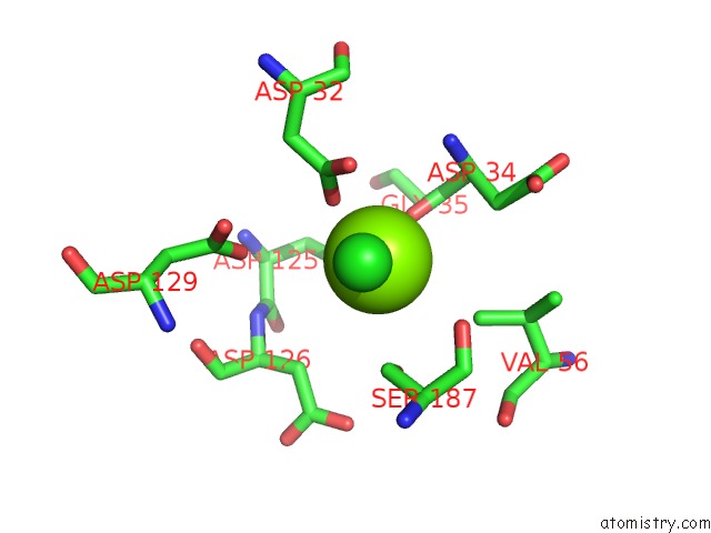
Mono view
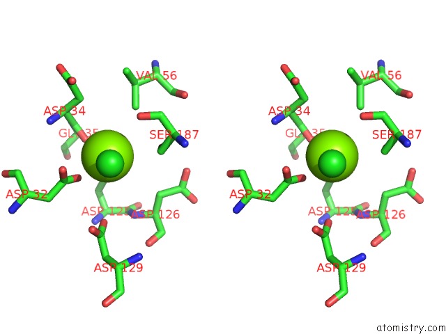
Stereo pair view
Mono view
Stereo pair view
A full contact list of Magnesium with other atoms in the Mg binding
site number 2 of Crystal Structure of E. Coli Phosphatase Yrbi, with Mg, Tetragonal Form within 5.0Å range:
|
Magnesium binding site 3 out of 8 in 3hyc
Go back to
Magnesium binding site 3 out
of 8 in the Crystal Structure of E. Coli Phosphatase Yrbi, with Mg, Tetragonal Form
Mono view
Stereo pair view
Mono view
Stereo pair view
A full contact list of Magnesium with other atoms in the Mg binding
site number 3 of Crystal Structure of E. Coli Phosphatase Yrbi, with Mg, Tetragonal Form within 5.0Å range:
|
Magnesium binding site 4 out of 8 in 3hyc
Go back to
Magnesium binding site 4 out
of 8 in the Crystal Structure of E. Coli Phosphatase Yrbi, with Mg, Tetragonal Form
Mono view
Stereo pair view
Mono view
Stereo pair view
A full contact list of Magnesium with other atoms in the Mg binding
site number 4 of Crystal Structure of E. Coli Phosphatase Yrbi, with Mg, Tetragonal Form within 5.0Å range:
|
Magnesium binding site 5 out of 8 in 3hyc
Go back to
Magnesium binding site 5 out
of 8 in the Crystal Structure of E. Coli Phosphatase Yrbi, with Mg, Tetragonal Form
Mono view
Stereo pair view
Mono view
Stereo pair view
A full contact list of Magnesium with other atoms in the Mg binding
site number 5 of Crystal Structure of E. Coli Phosphatase Yrbi, with Mg, Tetragonal Form within 5.0Å range:
|
Magnesium binding site 6 out of 8 in 3hyc
Go back to
Magnesium binding site 6 out
of 8 in the Crystal Structure of E. Coli Phosphatase Yrbi, with Mg, Tetragonal Form
Mono view
Stereo pair view
Mono view
Stereo pair view
A full contact list of Magnesium with other atoms in the Mg binding
site number 6 of Crystal Structure of E. Coli Phosphatase Yrbi, with Mg, Tetragonal Form within 5.0Å range:
|
Magnesium binding site 7 out of 8 in 3hyc
Go back to
Magnesium binding site 7 out
of 8 in the Crystal Structure of E. Coli Phosphatase Yrbi, with Mg, Tetragonal Form
Mono view
Stereo pair view
Mono view
Stereo pair view
A full contact list of Magnesium with other atoms in the Mg binding
site number 7 of Crystal Structure of E. Coli Phosphatase Yrbi, with Mg, Tetragonal Form within 5.0Å range:
|
Magnesium binding site 8 out of 8 in 3hyc
Go back to
Magnesium binding site 8 out
of 8 in the Crystal Structure of E. Coli Phosphatase Yrbi, with Mg, Tetragonal Form
Mono view
Stereo pair view
Mono view
Stereo pair view
A full contact list of Magnesium with other atoms in the Mg binding
site number 8 of Crystal Structure of E. Coli Phosphatase Yrbi, with Mg, Tetragonal Form within 5.0Å range:
|
Reference:
T.Biswas,
L.Yi,
P.Aggarwal,
J.Wu,
J.R.Rubin,
J.A.Stuckey,
R.W.Woodard,
O.V.Tsodikov.
The Tail of Kdsc: Conformational Changes Control the Activity of A Haloacid Dehalogenase Superfamily Phosphatase. J.Biol.Chem. V. 284 30594 2009.
ISSN: ISSN 0021-9258
PubMed: 19726684
DOI: 10.1074/JBC.M109.012278
Page generated: Wed Aug 14 15:30:15 2024
ISSN: ISSN 0021-9258
PubMed: 19726684
DOI: 10.1074/JBC.M109.012278
Last articles
Zn in 9MJ5Zn in 9HNW
Zn in 9G0L
Zn in 9FNE
Zn in 9DZN
Zn in 9E0I
Zn in 9D32
Zn in 9DAK
Zn in 8ZXC
Zn in 8ZUF