Magnesium »
PDB 3hx0-3i5x »
3hzh »
Magnesium in PDB 3hzh: Crystal Structure of the Chex-Chey-BEF3-Mg+2 Complex From Borrelia Burgdorferi
Protein crystallography data
The structure of Crystal Structure of the Chex-Chey-BEF3-Mg+2 Complex From Borrelia Burgdorferi, PDB code: 3hzh
was solved by
Y.Pazy,
R.E.Silversmith,
M.Guarinari,
R.Zhao,
with X-Ray Crystallography technique. A brief refinement statistics is given in the table below:
| Resolution Low / High (Å) | 29.64 / 1.96 |
| Space group | P 32 2 1 |
| Cell size a, b, c (Å), α, β, γ (°) | 63.705, 63.705, 175.685, 90.00, 90.00, 120.00 |
| R / Rfree (%) | 24.8 / 25.9 |
Other elements in 3hzh:
The structure of Crystal Structure of the Chex-Chey-BEF3-Mg+2 Complex From Borrelia Burgdorferi also contains other interesting chemical elements:
| Fluorine | (F) | 3 atoms |
Magnesium Binding Sites:
The binding sites of Magnesium atom in the Crystal Structure of the Chex-Chey-BEF3-Mg+2 Complex From Borrelia Burgdorferi
(pdb code 3hzh). This binding sites where shown within
5.0 Angstroms radius around Magnesium atom.
In total only one binding site of Magnesium was determined in the Crystal Structure of the Chex-Chey-BEF3-Mg+2 Complex From Borrelia Burgdorferi, PDB code: 3hzh:
In total only one binding site of Magnesium was determined in the Crystal Structure of the Chex-Chey-BEF3-Mg+2 Complex From Borrelia Burgdorferi, PDB code: 3hzh:
Magnesium binding site 1 out of 1 in 3hzh
Go back to
Magnesium binding site 1 out
of 1 in the Crystal Structure of the Chex-Chey-BEF3-Mg+2 Complex From Borrelia Burgdorferi
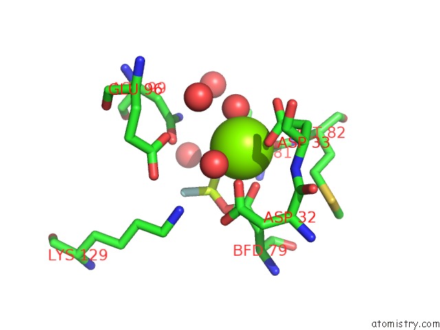
Mono view
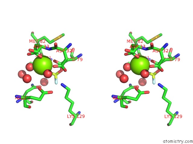
Stereo pair view
Mono view
Stereo pair view
A full contact list of Magnesium with other atoms in the Mg binding
site number 1 of Crystal Structure of the Chex-Chey-BEF3-Mg+2 Complex From Borrelia Burgdorferi within 5.0Å range:
|
Reference:
Y.Pazy,
M.A.Motaleb,
M.T.Guarnieri,
N.W.Charon,
R.Zhao,
R.E.Silversmith.
Identical Phosphatase Mechanisms Achieved Through Distinct Modes of Binding Phosphoprotein Substrate. Proc.Natl.Acad.Sci.Usa V. 107 1924 2010.
ISSN: ISSN 0027-8424
PubMed: 20080618
DOI: 10.1073/PNAS.0911185107
Page generated: Wed Aug 14 15:32:15 2024
ISSN: ISSN 0027-8424
PubMed: 20080618
DOI: 10.1073/PNAS.0911185107
Last articles
Zn in 9MJ5Zn in 9HNW
Zn in 9G0L
Zn in 9FNE
Zn in 9DZN
Zn in 9E0I
Zn in 9D32
Zn in 9DAK
Zn in 8ZXC
Zn in 8ZUF