Magnesium »
PDB 3hx0-3i5x »
3hzt »
Magnesium in PDB 3hzt: Crystal Structure of Toxoplasma Gondii CDPK3, TGME49_105860
Enzymatic activity of Crystal Structure of Toxoplasma Gondii CDPK3, TGME49_105860
All present enzymatic activity of Crystal Structure of Toxoplasma Gondii CDPK3, TGME49_105860:
2.7.11.17;
2.7.11.17;
Protein crystallography data
The structure of Crystal Structure of Toxoplasma Gondii CDPK3, TGME49_105860, PDB code: 3hzt
was solved by
A.K.Wernimont,
J.D.Artz,
P.Finnerty,
G.Wasney,
A.Allali-Hassani,
M.Vedadi,
A.Bochkarev,
C.H.Arrowsmith,
A.M.Edwards,
C.Bountra,
J.Weigelt,
R.Hui,
M.Amani,
Structural Genomics Consortium (Sgc),
with X-Ray Crystallography technique. A brief refinement statistics is given in the table below:
| Resolution Low / High (Å) | 30.00 / 2.00 |
| Space group | P 1 21 1 |
| Cell size a, b, c (Å), α, β, γ (°) | 71.051, 43.761, 84.821, 90.00, 96.95, 90.00 |
| R / Rfree (%) | 20.7 / 25.2 |
Other elements in 3hzt:
The structure of Crystal Structure of Toxoplasma Gondii CDPK3, TGME49_105860 also contains other interesting chemical elements:
| Chlorine | (Cl) | 1 atom |
Magnesium Binding Sites:
The binding sites of Magnesium atom in the Crystal Structure of Toxoplasma Gondii CDPK3, TGME49_105860
(pdb code 3hzt). This binding sites where shown within
5.0 Angstroms radius around Magnesium atom.
In total only one binding site of Magnesium was determined in the Crystal Structure of Toxoplasma Gondii CDPK3, TGME49_105860, PDB code: 3hzt:
In total only one binding site of Magnesium was determined in the Crystal Structure of Toxoplasma Gondii CDPK3, TGME49_105860, PDB code: 3hzt:
Magnesium binding site 1 out of 1 in 3hzt
Go back to
Magnesium binding site 1 out
of 1 in the Crystal Structure of Toxoplasma Gondii CDPK3, TGME49_105860
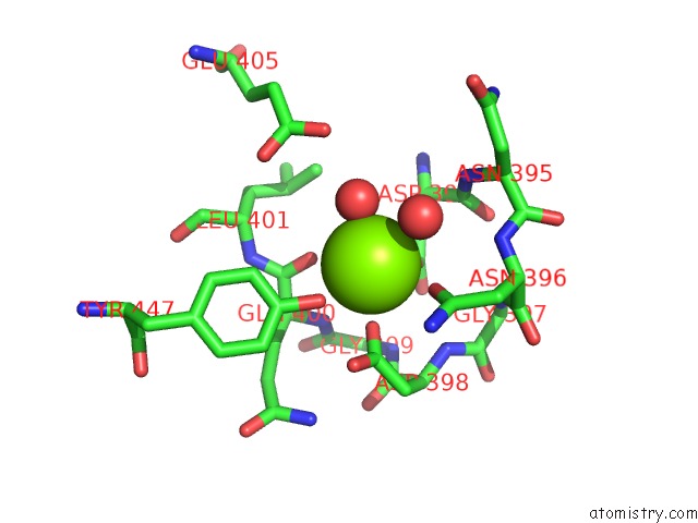
Mono view
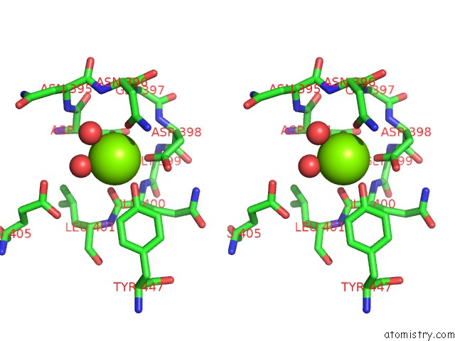
Stereo pair view
Mono view
Stereo pair view
A full contact list of Magnesium with other atoms in the Mg binding
site number 1 of Crystal Structure of Toxoplasma Gondii CDPK3, TGME49_105860 within 5.0Å range:
|
Reference:
A.K.Wernimont,
J.D.Artz,
P.Finerty,
Y.H.Lin,
M.Amani,
A.Allali-Hassani,
G.Senisterra,
M.Vedadi,
W.Tempel,
F.Mackenzie,
I.Chau,
S.Lourido,
L.D.Sibley,
R.Hui.
Structures of Apicomplexan Calcium-Dependent Protein Kinases Reveal Mechanism of Activation By Calcium. Nat.Struct.Mol.Biol. V. 17 596 2010.
ISSN: ISSN 1545-9993
PubMed: 20436473
DOI: 10.1038/NSMB.1795
Page generated: Wed Aug 14 15:32:49 2024
ISSN: ISSN 1545-9993
PubMed: 20436473
DOI: 10.1038/NSMB.1795
Last articles
Zn in 9MJ5Zn in 9HNW
Zn in 9G0L
Zn in 9FNE
Zn in 9DZN
Zn in 9E0I
Zn in 9D32
Zn in 9DAK
Zn in 8ZXC
Zn in 8ZUF