Magnesium »
PDB 3hx0-3i5x »
3i48 »
Magnesium in PDB 3i48: Crystal Structure of Beta Toxin From Staphylococcus Aureus F277A, P278A Mutant with Bound Magnesium Ions
Protein crystallography data
The structure of Crystal Structure of Beta Toxin From Staphylococcus Aureus F277A, P278A Mutant with Bound Magnesium Ions, PDB code: 3i48
was solved by
M.Huseby,
K.Shi,
A.C.Kruse,
D.H.Ohlendorf,
with X-Ray Crystallography technique. A brief refinement statistics is given in the table below:
| Resolution Low / High (Å) | 28.16 / 1.80 |
| Space group | P 21 21 21 |
| Cell size a, b, c (Å), α, β, γ (°) | 62.900, 68.577, 126.559, 90.00, 90.00, 90.00 |
| R / Rfree (%) | 18.4 / 22 |
Magnesium Binding Sites:
The binding sites of Magnesium atom in the Crystal Structure of Beta Toxin From Staphylococcus Aureus F277A, P278A Mutant with Bound Magnesium Ions
(pdb code 3i48). This binding sites where shown within
5.0 Angstroms radius around Magnesium atom.
In total 2 binding sites of Magnesium where determined in the Crystal Structure of Beta Toxin From Staphylococcus Aureus F277A, P278A Mutant with Bound Magnesium Ions, PDB code: 3i48:
Jump to Magnesium binding site number: 1; 2;
In total 2 binding sites of Magnesium where determined in the Crystal Structure of Beta Toxin From Staphylococcus Aureus F277A, P278A Mutant with Bound Magnesium Ions, PDB code: 3i48:
Jump to Magnesium binding site number: 1; 2;
Magnesium binding site 1 out of 2 in 3i48
Go back to
Magnesium binding site 1 out
of 2 in the Crystal Structure of Beta Toxin From Staphylococcus Aureus F277A, P278A Mutant with Bound Magnesium Ions
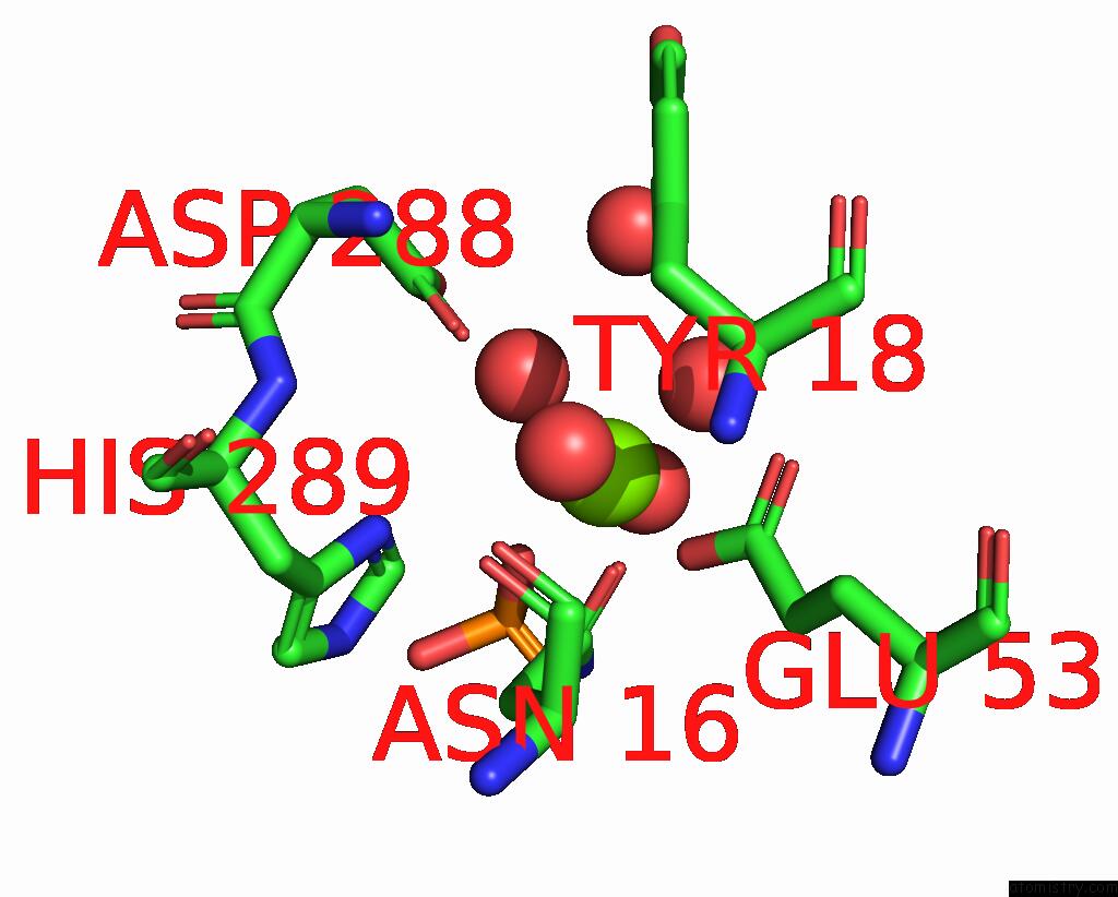
Mono view
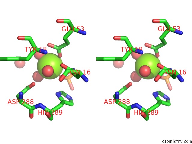
Stereo pair view
Mono view
Stereo pair view
A full contact list of Magnesium with other atoms in the Mg binding
site number 1 of Crystal Structure of Beta Toxin From Staphylococcus Aureus F277A, P278A Mutant with Bound Magnesium Ions within 5.0Å range:
|
Magnesium binding site 2 out of 2 in 3i48
Go back to
Magnesium binding site 2 out
of 2 in the Crystal Structure of Beta Toxin From Staphylococcus Aureus F277A, P278A Mutant with Bound Magnesium Ions
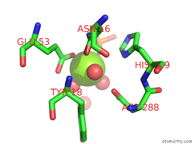
Mono view
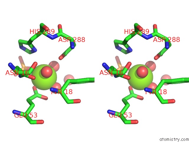
Stereo pair view
Mono view
Stereo pair view
A full contact list of Magnesium with other atoms in the Mg binding
site number 2 of Crystal Structure of Beta Toxin From Staphylococcus Aureus F277A, P278A Mutant with Bound Magnesium Ions within 5.0Å range:
|
Reference:
M.Huseby,
K.Shi,
A.C.Kruse,
J.Digre,
F.Mengistu,
G.A.Bohach,
P.S.Schlievert,
D.H.Ohlendorf,
C.A.Earhart.
Structure and Biological Functions of Beta Toxin From Staphylococcus Aureus: Role of the Hydrophobic Beta Hairpin in Virulence To Be Published 2009.
Page generated: Sun Aug 10 22:15:45 2025
Last articles
Mg in 4DV3Mg in 4DV2
Mg in 4DV0
Mg in 4DV1
Mg in 4DUZ
Mg in 4DUY
Mg in 4DR7
Mg in 4DR6
Mg in 4DR5
Mg in 4DUX