Magnesium »
PDB 3hx0-3i5x »
3i5f »
Magnesium in PDB 3i5f: Crystal Structure of Squid Mg.Adp Myosin S1
Protein crystallography data
The structure of Crystal Structure of Squid Mg.Adp Myosin S1, PDB code: 3i5f
was solved by
Y.Yang,
S.Gourinath,
M.Kovacs,
L.Nyitray,
R.Reutzel,
D.M.Himmel,
E.O'neall-Hennessey,
L.Reshetnikova,
A.G.Szent-Gyorgyi,
J.H.Brown,
C.Cohen,
with X-Ray Crystallography technique. A brief refinement statistics is given in the table below:
| Resolution Low / High (Å) | 43.23 / 3.10 |
| Space group | C 1 2 1 |
| Cell size a, b, c (Å), α, β, γ (°) | 187.415, 101.687, 94.751, 90.00, 119.75, 90.00 |
| R / Rfree (%) | 24.4 / 31.7 |
Magnesium Binding Sites:
The binding sites of Magnesium atom in the Crystal Structure of Squid Mg.Adp Myosin S1
(pdb code 3i5f). This binding sites where shown within
5.0 Angstroms radius around Magnesium atom.
In total only one binding site of Magnesium was determined in the Crystal Structure of Squid Mg.Adp Myosin S1, PDB code: 3i5f:
In total only one binding site of Magnesium was determined in the Crystal Structure of Squid Mg.Adp Myosin S1, PDB code: 3i5f:
Magnesium binding site 1 out of 1 in 3i5f
Go back to
Magnesium binding site 1 out
of 1 in the Crystal Structure of Squid Mg.Adp Myosin S1
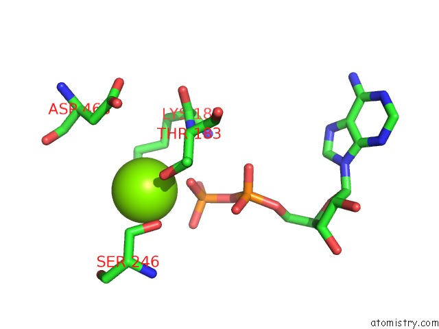
Mono view
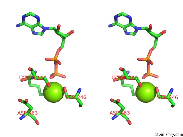
Stereo pair view
Mono view
Stereo pair view
A full contact list of Magnesium with other atoms in the Mg binding
site number 1 of Crystal Structure of Squid Mg.Adp Myosin S1 within 5.0Å range:
|
Reference:
Y.Yang,
S.Gourinath,
M.Kovacs,
L.Nyitray,
R.Reutzel,
D.M.Himmel,
E.O'neall-Hennessey,
L.Reshetnikova,
A.G.Szent-Gyorgyi,
J.H.Brown,
C.Cohen.
Rigor-Like Structures From Muscle Myosins Reveal Key Mechanical Elements in the Transduction Pathways of This Allosteric Motor. Structure V. 15 553 2007.
ISSN: ISSN 0969-2126
PubMed: 17502101
DOI: 10.1016/J.STR.2007.03.010
Page generated: Wed Aug 14 15:40:07 2024
ISSN: ISSN 0969-2126
PubMed: 17502101
DOI: 10.1016/J.STR.2007.03.010
Last articles
Zn in 9MJ5Zn in 9HNW
Zn in 9G0L
Zn in 9FNE
Zn in 9DZN
Zn in 9E0I
Zn in 9D32
Zn in 9DAK
Zn in 8ZXC
Zn in 8ZUF