Magnesium »
PDB 3i5y-3ig8 »
3i5y »
Magnesium in PDB 3i5y: Structure of MSS116P Bound to Ssrna Containing A Single 5-Bru and Amp- Pnp
Protein crystallography data
The structure of Structure of MSS116P Bound to Ssrna Containing A Single 5-Bru and Amp- Pnp, PDB code: 3i5y
was solved by
M.Del Campo,
A.M.Lambowitz,
with X-Ray Crystallography technique. A brief refinement statistics is given in the table below:
| Resolution Low / High (Å) | 34.88 / 2.49 |
| Space group | P 21 21 2 |
| Cell size a, b, c (Å), α, β, γ (°) | 89.190, 126.352, 55.990, 90.00, 90.00, 90.00 |
| R / Rfree (%) | 20.4 / 24.2 |
Other elements in 3i5y:
The structure of Structure of MSS116P Bound to Ssrna Containing A Single 5-Bru and Amp- Pnp also contains other interesting chemical elements:
| Bromine | (Br) | 1 atom |
Magnesium Binding Sites:
The binding sites of Magnesium atom in the Structure of MSS116P Bound to Ssrna Containing A Single 5-Bru and Amp- Pnp
(pdb code 3i5y). This binding sites where shown within
5.0 Angstroms radius around Magnesium atom.
In total only one binding site of Magnesium was determined in the Structure of MSS116P Bound to Ssrna Containing A Single 5-Bru and Amp- Pnp, PDB code: 3i5y:
In total only one binding site of Magnesium was determined in the Structure of MSS116P Bound to Ssrna Containing A Single 5-Bru and Amp- Pnp, PDB code: 3i5y:
Magnesium binding site 1 out of 1 in 3i5y
Go back to
Magnesium binding site 1 out
of 1 in the Structure of MSS116P Bound to Ssrna Containing A Single 5-Bru and Amp- Pnp
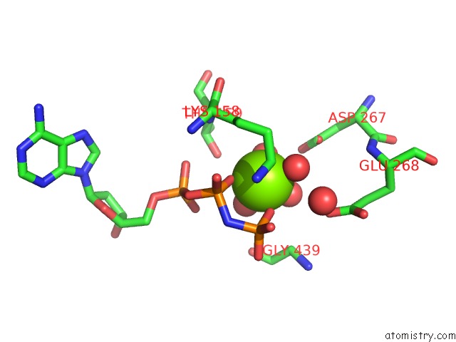
Mono view
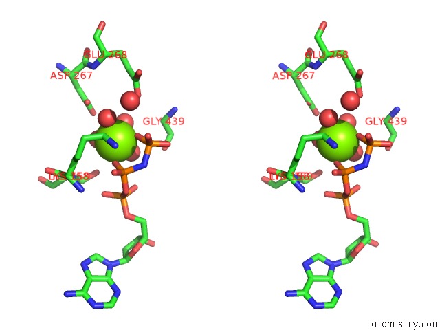
Stereo pair view
Mono view
Stereo pair view
A full contact list of Magnesium with other atoms in the Mg binding
site number 1 of Structure of MSS116P Bound to Ssrna Containing A Single 5-Bru and Amp- Pnp within 5.0Å range:
|
Reference:
M.Del Campo,
A.M.Lambowitz.
Structure of the Yeast Dead Box Protein MSS116P Reveals Two Wedges That Crimp Rna Mol.Cell V. 35 598 2009.
ISSN: ISSN 1097-2765
PubMed: 19748356
DOI: 10.1016/J.MOLCEL.2009.07.032
Page generated: Wed Aug 14 15:58:15 2024
ISSN: ISSN 1097-2765
PubMed: 19748356
DOI: 10.1016/J.MOLCEL.2009.07.032
Last articles
Fe in 2YXOFe in 2YRS
Fe in 2YXC
Fe in 2YNM
Fe in 2YVJ
Fe in 2YP1
Fe in 2YU2
Fe in 2YU1
Fe in 2YQB
Fe in 2YOO