Magnesium »
PDB 3i5y-3ig8 »
3i92 »
Magnesium in PDB 3i92: Structure of the Cytosolic Domain of E. Coli Feob, GPPCH2P-Bound Form
Protein crystallography data
The structure of Structure of the Cytosolic Domain of E. Coli Feob, GPPCH2P-Bound Form, PDB code: 3i92
was solved by
N.Petermann,
G.Hansen,
T.Hogg,
R.Hilgenfeld,
with X-Ray Crystallography technique. A brief refinement statistics is given in the table below:
| Resolution Low / High (Å) | 45.45 / 3.00 |
| Space group | P 1 21 1 |
| Cell size a, b, c (Å), α, β, γ (°) | 74.724, 55.925, 90.931, 90.00, 92.09, 90.00 |
| R / Rfree (%) | 21.7 / 27.2 |
Magnesium Binding Sites:
The binding sites of Magnesium atom in the Structure of the Cytosolic Domain of E. Coli Feob, GPPCH2P-Bound Form
(pdb code 3i92). This binding sites where shown within
5.0 Angstroms radius around Magnesium atom.
In total 3 binding sites of Magnesium where determined in the Structure of the Cytosolic Domain of E. Coli Feob, GPPCH2P-Bound Form, PDB code: 3i92:
Jump to Magnesium binding site number: 1; 2; 3;
In total 3 binding sites of Magnesium where determined in the Structure of the Cytosolic Domain of E. Coli Feob, GPPCH2P-Bound Form, PDB code: 3i92:
Jump to Magnesium binding site number: 1; 2; 3;
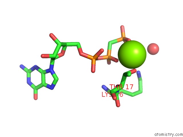
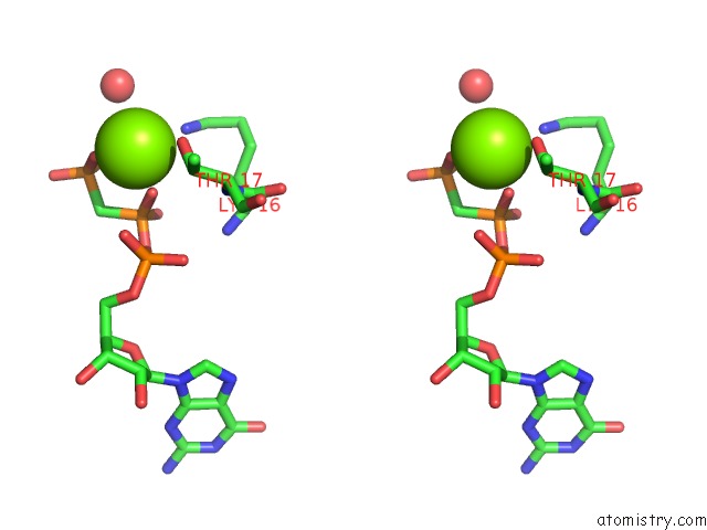
Magnesium binding site 1 out of 3 in 3i92
Go back to
Magnesium binding site 1 out
of 3 in the Structure of the Cytosolic Domain of E. Coli Feob, GPPCH2P-Bound Form
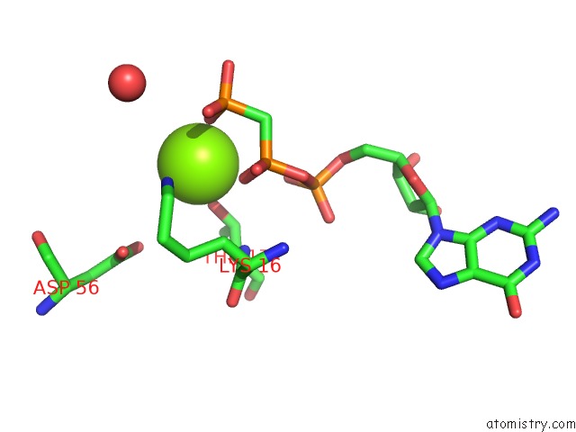
Mono view
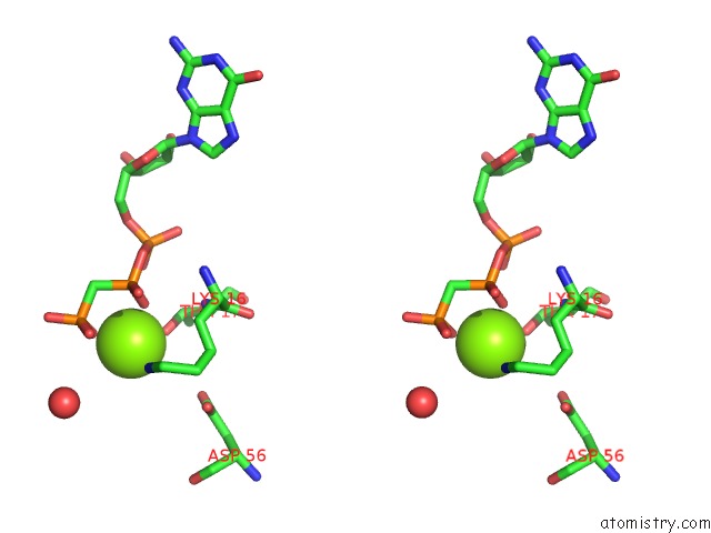
Stereo pair view
Mono view
Stereo pair view
A full contact list of Magnesium with other atoms in the Mg binding
site number 1 of Structure of the Cytosolic Domain of E. Coli Feob, GPPCH2P-Bound Form within 5.0Å range:
|
Magnesium binding site 2 out of 3 in 3i92
Go back to
Magnesium binding site 2 out
of 3 in the Structure of the Cytosolic Domain of E. Coli Feob, GPPCH2P-Bound Form
Mono view
Stereo pair view
Mono view
Stereo pair view
A full contact list of Magnesium with other atoms in the Mg binding
site number 2 of Structure of the Cytosolic Domain of E. Coli Feob, GPPCH2P-Bound Form within 5.0Å range:
|
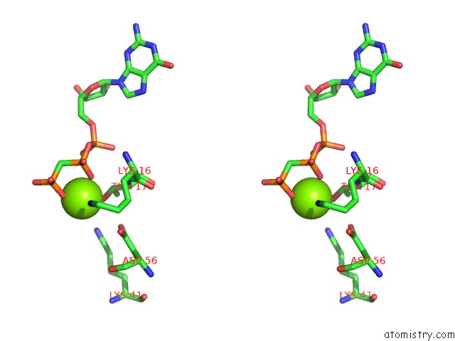
Magnesium binding site 3 out of 3 in 3i92
Go back to
Magnesium binding site 3 out
of 3 in the Structure of the Cytosolic Domain of E. Coli Feob, GPPCH2P-Bound Form
Mono view
Stereo pair view
Mono view
Stereo pair view
A full contact list of Magnesium with other atoms in the Mg binding
site number 3 of Structure of the Cytosolic Domain of E. Coli Feob, GPPCH2P-Bound Form within 5.0Å range:
|
Reference:
N.Petermann,
C.L.Schmidt,
G.Hansen,
A.K.Wagner,
R.Hilgenfeld,
T.Hogg.
Structural Basis For the Intrinsic Gtpase and Gdi Activities of Feob, A Prokaryotic Transmembrane Gtp/Gdp-Binding Protein To Be Published.
Page generated: Sun Aug 10 22:29:47 2025
Last articles
Mg in 4DV3Mg in 4DV2
Mg in 4DV0
Mg in 4DV1
Mg in 4DUZ
Mg in 4DUY
Mg in 4DR7
Mg in 4DR6
Mg in 4DR5
Mg in 4DUX