Magnesium »
PDB 3igi-3is5 »
3ii9 »
Magnesium in PDB 3ii9: Crystal Structure of Glutaryl-Coa Dehydrogenase From Burkholderia Pseudomallei at 1.73 Angstrom
Enzymatic activity of Crystal Structure of Glutaryl-Coa Dehydrogenase From Burkholderia Pseudomallei at 1.73 Angstrom
All present enzymatic activity of Crystal Structure of Glutaryl-Coa Dehydrogenase From Burkholderia Pseudomallei at 1.73 Angstrom:
1.3.99.7;
1.3.99.7;
Protein crystallography data
The structure of Crystal Structure of Glutaryl-Coa Dehydrogenase From Burkholderia Pseudomallei at 1.73 Angstrom, PDB code: 3ii9
was solved by
R.F.Ismagilov,
L.Li,
W.B.Du,
B.Staker,
Accelerated Technologies Centerfor Gene To 3D Structure (Atcg3D),
Seattle Structural Genomics Centerfor Infectious Disease (Ssgcid),
with X-Ray Crystallography technique. A brief refinement statistics is given in the table below:
| Resolution Low / High (Å) | 40.23 / 1.74 |
| Space group | P 1 21 1 |
| Cell size a, b, c (Å), α, β, γ (°) | 80.966, 141.268, 84.012, 90.00, 112.26, 90.00 |
| R / Rfree (%) | 17 / 19.6 |
Magnesium Binding Sites:
The binding sites of Magnesium atom in the Crystal Structure of Glutaryl-Coa Dehydrogenase From Burkholderia Pseudomallei at 1.73 Angstrom
(pdb code 3ii9). This binding sites where shown within
5.0 Angstroms radius around Magnesium atom.
In total 4 binding sites of Magnesium where determined in the Crystal Structure of Glutaryl-Coa Dehydrogenase From Burkholderia Pseudomallei at 1.73 Angstrom, PDB code: 3ii9:
Jump to Magnesium binding site number: 1; 2; 3; 4;
In total 4 binding sites of Magnesium where determined in the Crystal Structure of Glutaryl-Coa Dehydrogenase From Burkholderia Pseudomallei at 1.73 Angstrom, PDB code: 3ii9:
Jump to Magnesium binding site number: 1; 2; 3; 4;
Magnesium binding site 1 out of 4 in 3ii9
Go back to
Magnesium binding site 1 out
of 4 in the Crystal Structure of Glutaryl-Coa Dehydrogenase From Burkholderia Pseudomallei at 1.73 Angstrom

Mono view
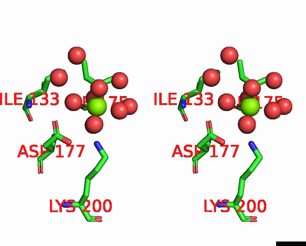
Stereo pair view
Mono view
Stereo pair view
A full contact list of Magnesium with other atoms in the Mg binding
site number 1 of Crystal Structure of Glutaryl-Coa Dehydrogenase From Burkholderia Pseudomallei at 1.73 Angstrom within 5.0Å range:
|
Magnesium binding site 2 out of 4 in 3ii9
Go back to
Magnesium binding site 2 out
of 4 in the Crystal Structure of Glutaryl-Coa Dehydrogenase From Burkholderia Pseudomallei at 1.73 Angstrom
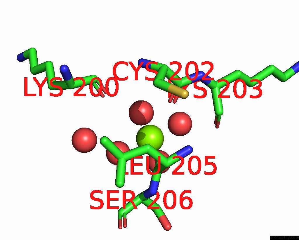
Mono view
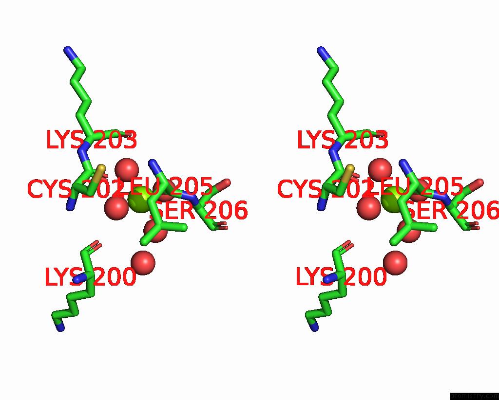
Stereo pair view
Mono view
Stereo pair view
A full contact list of Magnesium with other atoms in the Mg binding
site number 2 of Crystal Structure of Glutaryl-Coa Dehydrogenase From Burkholderia Pseudomallei at 1.73 Angstrom within 5.0Å range:
|
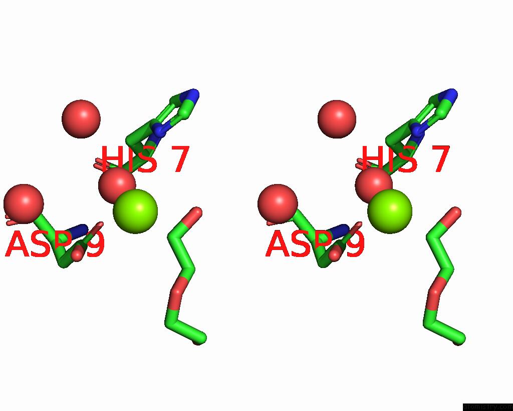
Magnesium binding site 3 out of 4 in 3ii9
Go back to
Magnesium binding site 3 out
of 4 in the Crystal Structure of Glutaryl-Coa Dehydrogenase From Burkholderia Pseudomallei at 1.73 Angstrom

Mono view

Stereo pair view
Mono view
Stereo pair view
A full contact list of Magnesium with other atoms in the Mg binding
site number 3 of Crystal Structure of Glutaryl-Coa Dehydrogenase From Burkholderia Pseudomallei at 1.73 Angstrom within 5.0Å range:
|
Magnesium binding site 4 out of 4 in 3ii9
Go back to
Magnesium binding site 4 out
of 4 in the Crystal Structure of Glutaryl-Coa Dehydrogenase From Burkholderia Pseudomallei at 1.73 Angstrom

Mono view
Stereo pair view
Mono view
Stereo pair view
A full contact list of Magnesium with other atoms in the Mg binding
site number 4 of Crystal Structure of Glutaryl-Coa Dehydrogenase From Burkholderia Pseudomallei at 1.73 Angstrom within 5.0Å range:
|
Reference:
L.Li,
W.Du,
R.Ismagilov.
User-Loaded Slipchip For Equipment-Free Multiplexed Nanoliter-Scale Experiments. J.Am.Chem.Soc. V. 132 106 2010.
ISSN: ISSN 0002-7863
PubMed: 20000708
DOI: 10.1021/JA908555N
Page generated: Sun Aug 10 22:35:18 2025
ISSN: ISSN 0002-7863
PubMed: 20000708
DOI: 10.1021/JA908555N
Last articles
Mg in 4OMFMg in 4OO1
Mg in 4OKM
Mg in 4OLS
Mg in 4OL0
Mg in 4OKK
Mg in 4OKJ
Mg in 4OKQ
Mg in 4OK9
Mg in 4OKE