Magnesium »
PDB 3igi-3is5 »
3iie »
Magnesium in PDB 3iie: 1-Deoxy-D-Xylulose 5-Phosphate Reductoisomerase From Yersinia Pestis.
Enzymatic activity of 1-Deoxy-D-Xylulose 5-Phosphate Reductoisomerase From Yersinia Pestis.
All present enzymatic activity of 1-Deoxy-D-Xylulose 5-Phosphate Reductoisomerase From Yersinia Pestis.:
1.1.1.267;
1.1.1.267;
Protein crystallography data
The structure of 1-Deoxy-D-Xylulose 5-Phosphate Reductoisomerase From Yersinia Pestis., PDB code: 3iie
was solved by
J.Osipiuk,
R.Mulligan,
J.Stam,
W.F.Anderson,
A.Joachimiak,
with X-Ray Crystallography technique. A brief refinement statistics is given in the table below:
| Resolution Low / High (Å) | 50.00 / 2.21 |
| Space group | P 32 2 1 |
| Cell size a, b, c (Å), α, β, γ (°) | 121.460, 121.460, 86.923, 90.00, 90.00, 120.00 |
| R / Rfree (%) | 17.9 / 24.1 |
Magnesium Binding Sites:
The binding sites of Magnesium atom in the 1-Deoxy-D-Xylulose 5-Phosphate Reductoisomerase From Yersinia Pestis.
(pdb code 3iie). This binding sites where shown within
5.0 Angstroms radius around Magnesium atom.
In total 3 binding sites of Magnesium where determined in the 1-Deoxy-D-Xylulose 5-Phosphate Reductoisomerase From Yersinia Pestis., PDB code: 3iie:
Jump to Magnesium binding site number: 1; 2; 3;
In total 3 binding sites of Magnesium where determined in the 1-Deoxy-D-Xylulose 5-Phosphate Reductoisomerase From Yersinia Pestis., PDB code: 3iie:
Jump to Magnesium binding site number: 1; 2; 3;
Magnesium binding site 1 out of 3 in 3iie
Go back to
Magnesium binding site 1 out
of 3 in the 1-Deoxy-D-Xylulose 5-Phosphate Reductoisomerase From Yersinia Pestis.
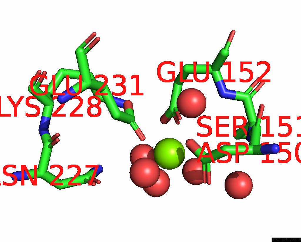
Mono view
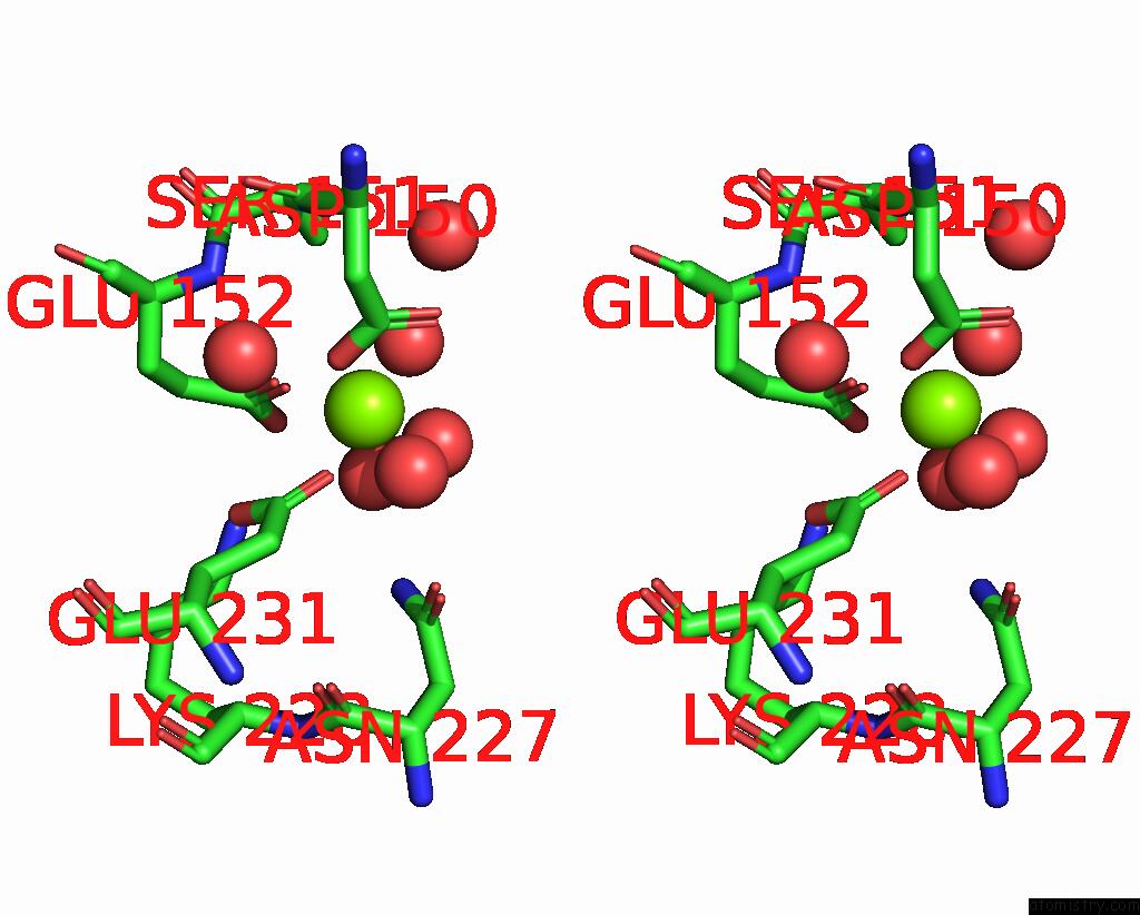
Stereo pair view
Mono view
Stereo pair view
A full contact list of Magnesium with other atoms in the Mg binding
site number 1 of 1-Deoxy-D-Xylulose 5-Phosphate Reductoisomerase From Yersinia Pestis. within 5.0Å range:
|
Magnesium binding site 2 out of 3 in 3iie
Go back to
Magnesium binding site 2 out
of 3 in the 1-Deoxy-D-Xylulose 5-Phosphate Reductoisomerase From Yersinia Pestis.

Mono view
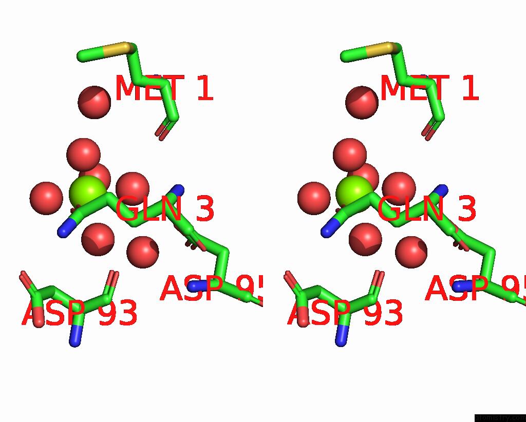
Stereo pair view
Mono view
Stereo pair view
A full contact list of Magnesium with other atoms in the Mg binding
site number 2 of 1-Deoxy-D-Xylulose 5-Phosphate Reductoisomerase From Yersinia Pestis. within 5.0Å range:
|
Magnesium binding site 3 out of 3 in 3iie
Go back to
Magnesium binding site 3 out
of 3 in the 1-Deoxy-D-Xylulose 5-Phosphate Reductoisomerase From Yersinia Pestis.

Mono view
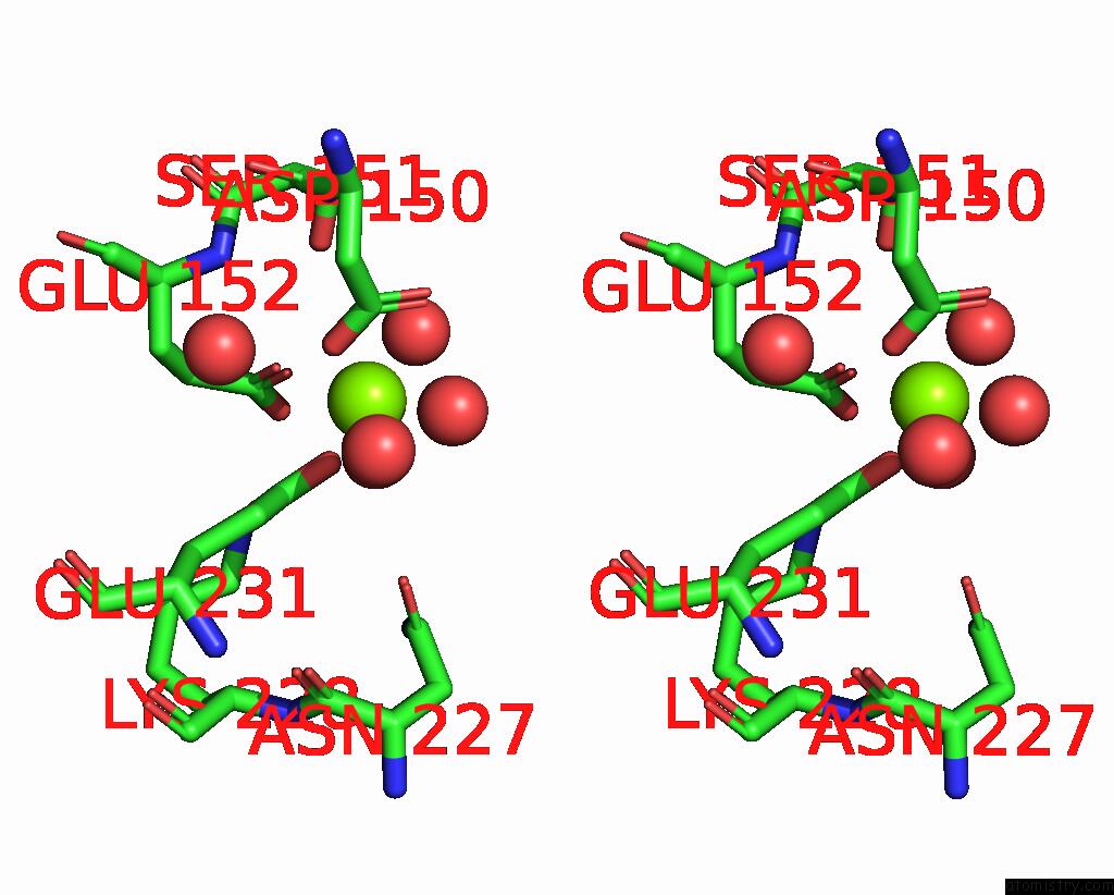
Stereo pair view
Mono view
Stereo pair view
A full contact list of Magnesium with other atoms in the Mg binding
site number 3 of 1-Deoxy-D-Xylulose 5-Phosphate Reductoisomerase From Yersinia Pestis. within 5.0Å range:
|
Reference:
J.Osipiuk,
R.Mulligan,
J.Stam,
W.F.Anderson,
A.Joachimiak.
X-Ray Crystal Structure of 1-Deoxy-D-Xylulose 5-Phosphate Reductoisomerase From Yersinia Pestis. To Be Published.
Page generated: Sun Aug 10 22:35:18 2025
Last articles
Mg in 4DV1Mg in 4DUZ
Mg in 4DUY
Mg in 4DR7
Mg in 4DR6
Mg in 4DR5
Mg in 4DUX
Mg in 4DUW
Mg in 4DUV
Mg in 4DUO