Magnesium »
PDB 3igi-3is5 »
3ild »
Magnesium in PDB 3ild: Structure of ORF157-K57A From Acidianus Filamentous Virus 1
Protein crystallography data
The structure of Structure of ORF157-K57A From Acidianus Filamentous Virus 1, PDB code: 3ild
was solved by
A.Goulet,
J.Lichiere,
D.Prangishvili,
H.Van Tilbeurgh,
C.Cambillau,
V.Campanacci,
with X-Ray Crystallography technique. A brief refinement statistics is given in the table below:
| Resolution Low / High (Å) | 22.00 / 3.10 |
| Space group | P 31 2 1 |
| Cell size a, b, c (Å), α, β, γ (°) | 64.757, 64.757, 85.743, 90.00, 90.00, 120.00 |
| R / Rfree (%) | 27.1 / 30.9 |
Magnesium Binding Sites:
The binding sites of Magnesium atom in the Structure of ORF157-K57A From Acidianus Filamentous Virus 1
(pdb code 3ild). This binding sites where shown within
5.0 Angstroms radius around Magnesium atom.
In total only one binding site of Magnesium was determined in the Structure of ORF157-K57A From Acidianus Filamentous Virus 1, PDB code: 3ild:
In total only one binding site of Magnesium was determined in the Structure of ORF157-K57A From Acidianus Filamentous Virus 1, PDB code: 3ild:
Magnesium binding site 1 out of 1 in 3ild
Go back to
Magnesium binding site 1 out
of 1 in the Structure of ORF157-K57A From Acidianus Filamentous Virus 1
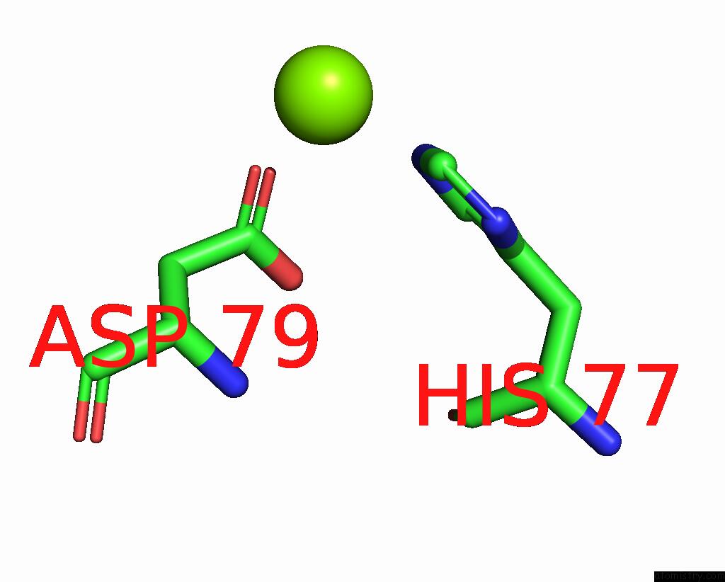
Mono view
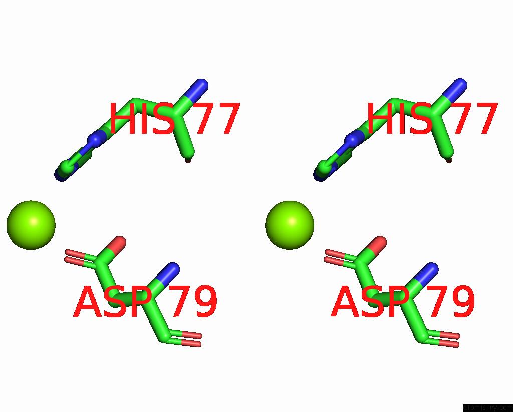
Stereo pair view
Mono view
Stereo pair view
A full contact list of Magnesium with other atoms in the Mg binding
site number 1 of Structure of ORF157-K57A From Acidianus Filamentous Virus 1 within 5.0Å range:
|
Reference:
A.Goulet,
M.Pina,
P.Redder,
D.Prangishvili,
L.Vera,
J.Lichiere,
N.Leulliot,
H.Van Tilbeurgh,
M.Ortiz-Lombardia,
V.Campanacci,
C.Cambillau.
ORF157 From the Archaeal Virus Acidianus Filamentous Virus 1 Defines A New Class of Nuclease J.Virol. V. 84 5025 2010.
ISSN: ISSN 0022-538X
PubMed: 20200253
DOI: 10.1128/JVI.01664-09
Page generated: Sun Aug 10 22:38:58 2025
ISSN: ISSN 0022-538X
PubMed: 20200253
DOI: 10.1128/JVI.01664-09
Last articles
Mg in 6HNQMg in 6HOS
Mg in 6HNS
Mg in 6HN2
Mg in 6HMZ
Mg in 6HMU
Mg in 6HMT
Mg in 6HLR
Mg in 6HLQ
Mg in 6HKY