Magnesium in PDB 3j7h: Structure of Beta-Galactosidase at 3.2-A Resolution Obtained By Cryo- Electron Microscopy
Enzymatic activity of Structure of Beta-Galactosidase at 3.2-A Resolution Obtained By Cryo- Electron Microscopy
All present enzymatic activity of Structure of Beta-Galactosidase at 3.2-A Resolution Obtained By Cryo- Electron Microscopy:
3.2.1.23;
Magnesium Binding Sites:
The binding sites of Magnesium atom in the Structure of Beta-Galactosidase at 3.2-A Resolution Obtained By Cryo- Electron Microscopy
(pdb code 3j7h). This binding sites where shown within
5.0 Angstroms radius around Magnesium atom.
In total 4 binding sites of Magnesium where determined in the
Structure of Beta-Galactosidase at 3.2-A Resolution Obtained By Cryo- Electron Microscopy, PDB code: 3j7h:
Jump to Magnesium binding site number:
1;
2;
3;
4;
Magnesium binding site 1 out
of 4 in 3j7h
Go back to
Magnesium Binding Sites List in 3j7h
Magnesium binding site 1 out
of 4 in the Structure of Beta-Galactosidase at 3.2-A Resolution Obtained By Cryo- Electron Microscopy
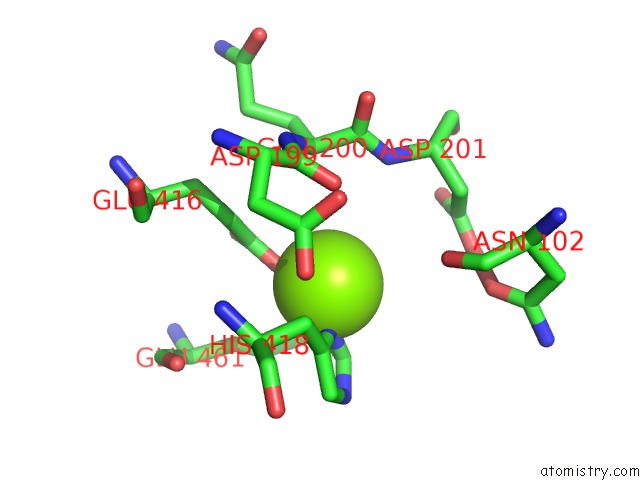
Mono view
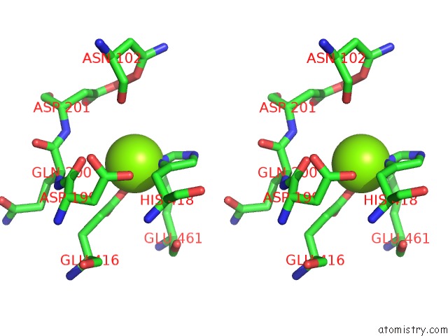
Stereo pair view
|
A full contact list of Magnesium with other atoms in the Mg binding
site number 1 of Structure of Beta-Galactosidase at 3.2-A Resolution Obtained By Cryo- Electron Microscopy within 5.0Å range:
probe
|
atom
|
residue
|
distance (Å)
|
B
|
Occ
|
A:Mg3001
b:16.1
occ:1.00
|
ND1
|
A:HIS418
|
1.5
|
11.4
|
1.0
|
OE2
|
A:GLU416
|
1.6
|
11.7
|
1.0
|
OE1
|
A:GLU461
|
1.7
|
13.9
|
1.0
|
CE1
|
A:HIS418
|
2.4
|
12.3
|
1.0
|
CG
|
A:HIS418
|
2.6
|
15.0
|
1.0
|
CD
|
A:GLU416
|
2.8
|
15.4
|
1.0
|
CD
|
A:GLU461
|
3.0
|
19.5
|
1.0
|
CB
|
A:HIS418
|
3.2
|
13.0
|
1.0
|
NE2
|
A:HIS418
|
3.5
|
11.3
|
1.0
|
CD2
|
A:HIS418
|
3.6
|
12.0
|
1.0
|
OE1
|
A:GLU416
|
3.7
|
11.8
|
1.0
|
OE2
|
A:GLU461
|
3.8
|
13.7
|
1.0
|
CG
|
A:GLU416
|
3.8
|
8.0
|
1.0
|
CB
|
A:GLU461
|
3.8
|
10.0
|
1.0
|
CG
|
A:GLU461
|
4.0
|
9.5
|
1.0
|
O
|
A:ASP199
|
4.2
|
15.8
|
1.0
|
O
|
A:ASN102
|
4.3
|
13.8
|
1.0
|
CA
|
A:GLN200
|
4.3
|
7.8
|
1.0
|
N
|
A:ASP201
|
4.4
|
10.8
|
1.0
|
CA
|
A:HIS418
|
4.4
|
12.2
|
1.0
|
C
|
A:ASP199
|
4.7
|
15.3
|
1.0
|
N
|
A:HIS418
|
4.7
|
13.6
|
1.0
|
CG
|
A:ASP201
|
4.8
|
17.0
|
1.0
|
C
|
A:GLN200
|
4.8
|
12.7
|
1.0
|
OD2
|
A:ASP201
|
4.9
|
11.7
|
1.0
|
N
|
A:GLN200
|
4.9
|
9.2
|
1.0
|
|
Magnesium binding site 2 out
of 4 in 3j7h
Go back to
Magnesium Binding Sites List in 3j7h
Magnesium binding site 2 out
of 4 in the Structure of Beta-Galactosidase at 3.2-A Resolution Obtained By Cryo- Electron Microscopy
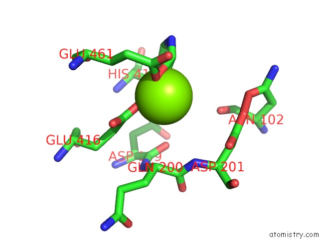
Mono view
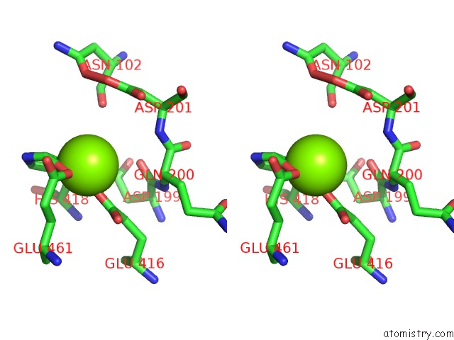
Stereo pair view
|
A full contact list of Magnesium with other atoms in the Mg binding
site number 2 of Structure of Beta-Galactosidase at 3.2-A Resolution Obtained By Cryo- Electron Microscopy within 5.0Å range:
probe
|
atom
|
residue
|
distance (Å)
|
B
|
Occ
|
B:Mg3001
b:16.1
occ:1.00
|
ND1
|
B:HIS418
|
1.5
|
11.4
|
1.0
|
OE2
|
B:GLU416
|
1.6
|
11.7
|
1.0
|
OE1
|
B:GLU461
|
1.7
|
13.9
|
1.0
|
CE1
|
B:HIS418
|
2.4
|
12.3
|
1.0
|
CG
|
B:HIS418
|
2.6
|
15.0
|
1.0
|
CD
|
B:GLU416
|
2.8
|
15.4
|
1.0
|
CD
|
B:GLU461
|
3.0
|
19.5
|
1.0
|
CB
|
B:HIS418
|
3.2
|
13.0
|
1.0
|
NE2
|
B:HIS418
|
3.5
|
11.3
|
1.0
|
CD2
|
B:HIS418
|
3.6
|
12.0
|
1.0
|
OE1
|
B:GLU416
|
3.7
|
11.8
|
1.0
|
OE2
|
B:GLU461
|
3.8
|
13.7
|
1.0
|
CG
|
B:GLU416
|
3.8
|
8.0
|
1.0
|
CB
|
B:GLU461
|
3.8
|
10.0
|
1.0
|
CG
|
B:GLU461
|
4.0
|
9.5
|
1.0
|
O
|
B:ASP199
|
4.2
|
15.8
|
1.0
|
O
|
B:ASN102
|
4.3
|
13.8
|
1.0
|
CA
|
B:GLN200
|
4.3
|
7.8
|
1.0
|
N
|
B:ASP201
|
4.4
|
10.8
|
1.0
|
CA
|
B:HIS418
|
4.4
|
12.2
|
1.0
|
C
|
B:ASP199
|
4.7
|
15.3
|
1.0
|
N
|
B:HIS418
|
4.7
|
13.6
|
1.0
|
CG
|
B:ASP201
|
4.8
|
17.0
|
1.0
|
C
|
B:GLN200
|
4.8
|
12.7
|
1.0
|
OD2
|
B:ASP201
|
4.9
|
11.7
|
1.0
|
N
|
B:GLN200
|
4.9
|
9.2
|
1.0
|
|
Magnesium binding site 3 out
of 4 in 3j7h
Go back to
Magnesium Binding Sites List in 3j7h
Magnesium binding site 3 out
of 4 in the Structure of Beta-Galactosidase at 3.2-A Resolution Obtained By Cryo- Electron Microscopy
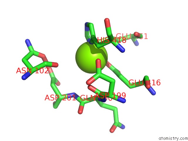
Mono view
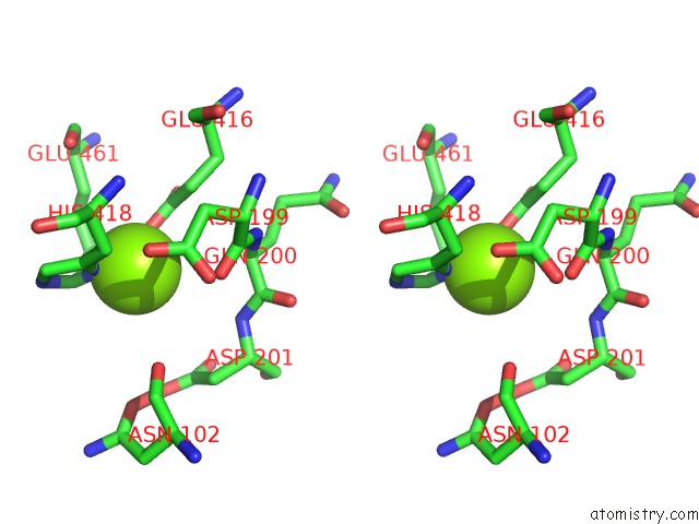
Stereo pair view
|
A full contact list of Magnesium with other atoms in the Mg binding
site number 3 of Structure of Beta-Galactosidase at 3.2-A Resolution Obtained By Cryo- Electron Microscopy within 5.0Å range:
probe
|
atom
|
residue
|
distance (Å)
|
B
|
Occ
|
C:Mg3001
b:16.1
occ:1.00
|
ND1
|
C:HIS418
|
1.5
|
11.4
|
1.0
|
OE2
|
C:GLU416
|
1.6
|
11.7
|
1.0
|
OE1
|
C:GLU461
|
1.7
|
13.9
|
1.0
|
CE1
|
C:HIS418
|
2.4
|
12.3
|
1.0
|
CG
|
C:HIS418
|
2.6
|
15.0
|
1.0
|
CD
|
C:GLU416
|
2.8
|
15.4
|
1.0
|
CD
|
C:GLU461
|
3.0
|
19.5
|
1.0
|
CB
|
C:HIS418
|
3.2
|
13.0
|
1.0
|
NE2
|
C:HIS418
|
3.5
|
11.3
|
1.0
|
CD2
|
C:HIS418
|
3.6
|
12.0
|
1.0
|
OE1
|
C:GLU416
|
3.7
|
11.8
|
1.0
|
OE2
|
C:GLU461
|
3.8
|
13.7
|
1.0
|
CG
|
C:GLU416
|
3.8
|
8.0
|
1.0
|
CB
|
C:GLU461
|
3.8
|
10.0
|
1.0
|
CG
|
C:GLU461
|
4.0
|
9.5
|
1.0
|
O
|
C:ASP199
|
4.2
|
15.8
|
1.0
|
O
|
C:ASN102
|
4.3
|
13.8
|
1.0
|
CA
|
C:GLN200
|
4.3
|
7.8
|
1.0
|
N
|
C:ASP201
|
4.4
|
10.8
|
1.0
|
CA
|
C:HIS418
|
4.4
|
12.2
|
1.0
|
C
|
C:ASP199
|
4.7
|
15.3
|
1.0
|
N
|
C:HIS418
|
4.7
|
13.6
|
1.0
|
CG
|
C:ASP201
|
4.8
|
17.0
|
1.0
|
C
|
C:GLN200
|
4.8
|
12.7
|
1.0
|
OD2
|
C:ASP201
|
4.9
|
11.7
|
1.0
|
N
|
C:GLN200
|
4.9
|
9.2
|
1.0
|
|
Magnesium binding site 4 out
of 4 in 3j7h
Go back to
Magnesium Binding Sites List in 3j7h
Magnesium binding site 4 out
of 4 in the Structure of Beta-Galactosidase at 3.2-A Resolution Obtained By Cryo- Electron Microscopy
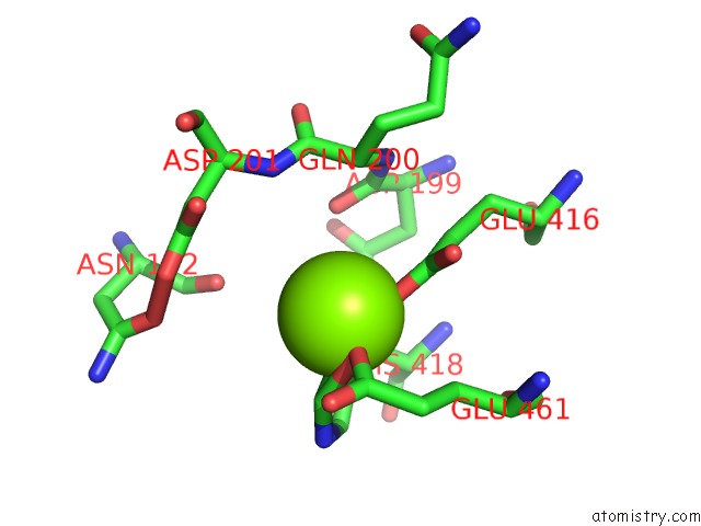
Mono view
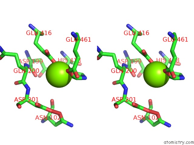
Stereo pair view
|
A full contact list of Magnesium with other atoms in the Mg binding
site number 4 of Structure of Beta-Galactosidase at 3.2-A Resolution Obtained By Cryo- Electron Microscopy within 5.0Å range:
probe
|
atom
|
residue
|
distance (Å)
|
B
|
Occ
|
D:Mg3001
b:16.1
occ:1.00
|
ND1
|
D:HIS418
|
1.5
|
11.4
|
1.0
|
OE2
|
D:GLU416
|
1.6
|
11.7
|
1.0
|
OE1
|
D:GLU461
|
1.7
|
13.9
|
1.0
|
CE1
|
D:HIS418
|
2.4
|
12.3
|
1.0
|
CG
|
D:HIS418
|
2.6
|
15.0
|
1.0
|
CD
|
D:GLU416
|
2.8
|
15.4
|
1.0
|
CD
|
D:GLU461
|
3.0
|
19.5
|
1.0
|
CB
|
D:HIS418
|
3.2
|
13.0
|
1.0
|
NE2
|
D:HIS418
|
3.5
|
11.3
|
1.0
|
CD2
|
D:HIS418
|
3.6
|
12.0
|
1.0
|
OE1
|
D:GLU416
|
3.7
|
11.8
|
1.0
|
OE2
|
D:GLU461
|
3.8
|
13.7
|
1.0
|
CG
|
D:GLU416
|
3.8
|
8.0
|
1.0
|
CB
|
D:GLU461
|
3.8
|
10.0
|
1.0
|
CG
|
D:GLU461
|
4.0
|
9.5
|
1.0
|
O
|
D:ASP199
|
4.2
|
15.8
|
1.0
|
O
|
D:ASN102
|
4.3
|
13.8
|
1.0
|
CA
|
D:GLN200
|
4.3
|
7.8
|
1.0
|
N
|
D:ASP201
|
4.4
|
10.8
|
1.0
|
CA
|
D:HIS418
|
4.4
|
12.2
|
1.0
|
C
|
D:ASP199
|
4.7
|
15.3
|
1.0
|
N
|
D:HIS418
|
4.7
|
13.6
|
1.0
|
CG
|
D:ASP201
|
4.8
|
17.0
|
1.0
|
C
|
D:GLN200
|
4.8
|
12.7
|
1.0
|
OD2
|
D:ASP201
|
4.9
|
11.7
|
1.0
|
N
|
D:GLN200
|
4.9
|
9.2
|
1.0
|
|
Reference:
A.Bartesaghi,
D.Matthies,
S.Banerjee,
A.Merk,
S.Subramaniam.
Structure of Beta-Galactosidase at 3.2- Angstrom Resolution Obtained By Cryo-Electron Microscopy. Proc.Natl.Acad.Sci.Usa V. 111 11709 2014.
ISSN: ISSN 0027-8424
PubMed: 25071206
DOI: 10.1073/PNAS.1402809111
Page generated: Wed Aug 14 16:25:53 2024
|