Magnesium »
PDB 3jay-3jwr »
3jr2 »
Magnesium in PDB 3jr2: X-Ray Crystal Structure of the Mg-Bound 3-Keto-L-Gulonate-6-Phosphate Decarboxylase From Vibrio Cholerae O1 Biovar El Tor Str. N16961
Protein crystallography data
The structure of X-Ray Crystal Structure of the Mg-Bound 3-Keto-L-Gulonate-6-Phosphate Decarboxylase From Vibrio Cholerae O1 Biovar El Tor Str. N16961, PDB code: 3jr2
was solved by
B.Nocek,
N.Maltseva,
J.Stam,
W.Anderson,
A.Joachimiak,
Csgid,
Center Forstructural Genomics Of Infectious Diseases (Csgid),
with X-Ray Crystallography technique. A brief refinement statistics is given in the table below:
| Resolution Low / High (Å) | 40.00 / 1.80 |
| Space group | P 21 21 21 |
| Cell size a, b, c (Å), α, β, γ (°) | 82.305, 95.711, 125.577, 90.00, 90.00, 90.00 |
| R / Rfree (%) | 16.3 / 20.2 |
Magnesium Binding Sites:
The binding sites of Magnesium atom in the X-Ray Crystal Structure of the Mg-Bound 3-Keto-L-Gulonate-6-Phosphate Decarboxylase From Vibrio Cholerae O1 Biovar El Tor Str. N16961
(pdb code 3jr2). This binding sites where shown within
5.0 Angstroms radius around Magnesium atom.
In total 7 binding sites of Magnesium where determined in the X-Ray Crystal Structure of the Mg-Bound 3-Keto-L-Gulonate-6-Phosphate Decarboxylase From Vibrio Cholerae O1 Biovar El Tor Str. N16961, PDB code: 3jr2:
Jump to Magnesium binding site number: 1; 2; 3; 4; 5; 6; 7;
In total 7 binding sites of Magnesium where determined in the X-Ray Crystal Structure of the Mg-Bound 3-Keto-L-Gulonate-6-Phosphate Decarboxylase From Vibrio Cholerae O1 Biovar El Tor Str. N16961, PDB code: 3jr2:
Jump to Magnesium binding site number: 1; 2; 3; 4; 5; 6; 7;
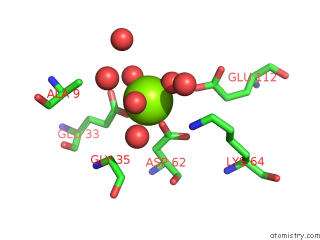
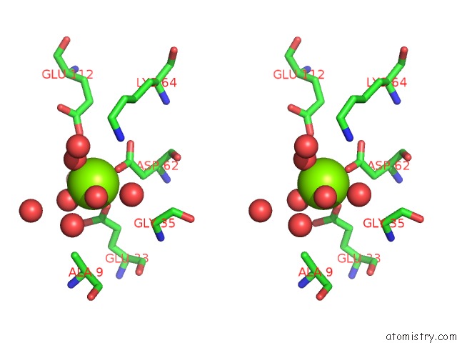
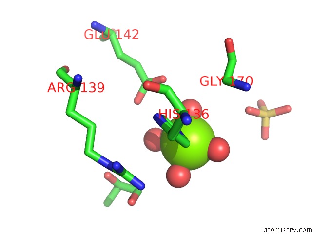
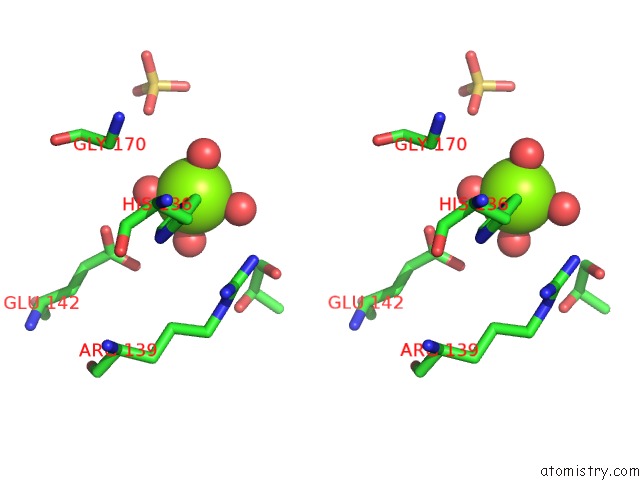
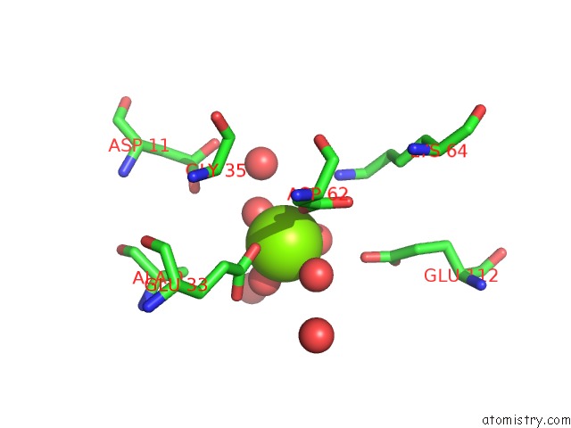
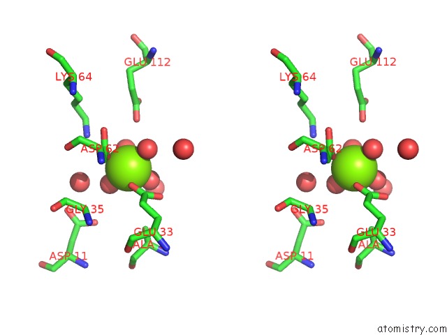
Magnesium binding site 1 out of 7 in 3jr2
Go back to
Magnesium binding site 1 out
of 7 in the X-Ray Crystal Structure of the Mg-Bound 3-Keto-L-Gulonate-6-Phosphate Decarboxylase From Vibrio Cholerae O1 Biovar El Tor Str. N16961
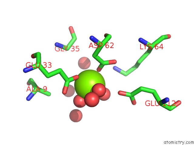
Mono view
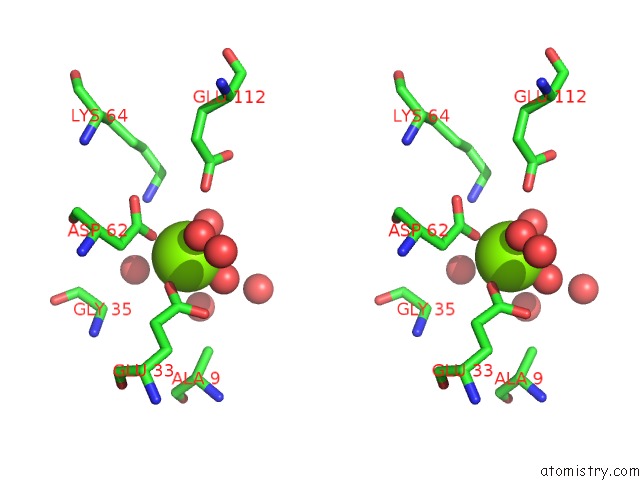
Stereo pair view
Mono view
Stereo pair view
A full contact list of Magnesium with other atoms in the Mg binding
site number 1 of X-Ray Crystal Structure of the Mg-Bound 3-Keto-L-Gulonate-6-Phosphate Decarboxylase From Vibrio Cholerae O1 Biovar El Tor Str. N16961 within 5.0Å range:
|
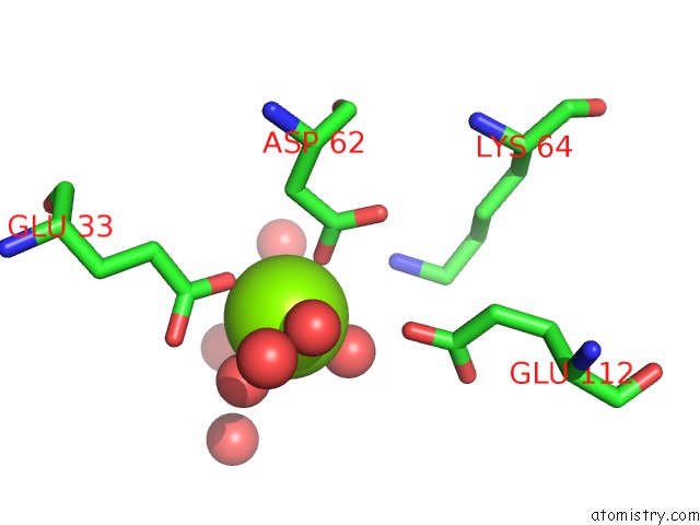
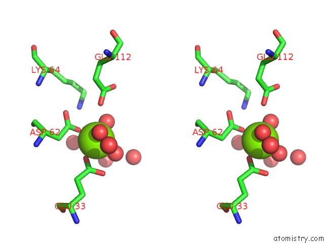
Magnesium binding site 2 out of 7 in 3jr2
Go back to
Magnesium binding site 2 out
of 7 in the X-Ray Crystal Structure of the Mg-Bound 3-Keto-L-Gulonate-6-Phosphate Decarboxylase From Vibrio Cholerae O1 Biovar El Tor Str. N16961
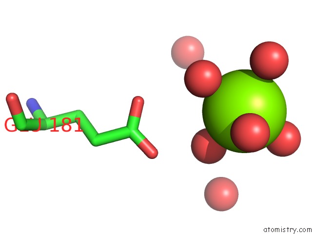
Mono view
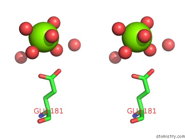
Stereo pair view
Mono view
Stereo pair view
A full contact list of Magnesium with other atoms in the Mg binding
site number 2 of X-Ray Crystal Structure of the Mg-Bound 3-Keto-L-Gulonate-6-Phosphate Decarboxylase From Vibrio Cholerae O1 Biovar El Tor Str. N16961 within 5.0Å range:
|
Magnesium binding site 3 out of 7 in 3jr2
Go back to
Magnesium binding site 3 out
of 7 in the X-Ray Crystal Structure of the Mg-Bound 3-Keto-L-Gulonate-6-Phosphate Decarboxylase From Vibrio Cholerae O1 Biovar El Tor Str. N16961
Mono view
Stereo pair view
Mono view
Stereo pair view
A full contact list of Magnesium with other atoms in the Mg binding
site number 3 of X-Ray Crystal Structure of the Mg-Bound 3-Keto-L-Gulonate-6-Phosphate Decarboxylase From Vibrio Cholerae O1 Biovar El Tor Str. N16961 within 5.0Å range:
|
Magnesium binding site 4 out of 7 in 3jr2
Go back to
Magnesium binding site 4 out
of 7 in the X-Ray Crystal Structure of the Mg-Bound 3-Keto-L-Gulonate-6-Phosphate Decarboxylase From Vibrio Cholerae O1 Biovar El Tor Str. N16961
Mono view
Stereo pair view
Mono view
Stereo pair view
A full contact list of Magnesium with other atoms in the Mg binding
site number 4 of X-Ray Crystal Structure of the Mg-Bound 3-Keto-L-Gulonate-6-Phosphate Decarboxylase From Vibrio Cholerae O1 Biovar El Tor Str. N16961 within 5.0Å range:
|
Magnesium binding site 5 out of 7 in 3jr2
Go back to
Magnesium binding site 5 out
of 7 in the X-Ray Crystal Structure of the Mg-Bound 3-Keto-L-Gulonate-6-Phosphate Decarboxylase From Vibrio Cholerae O1 Biovar El Tor Str. N16961
Mono view
Stereo pair view
Mono view
Stereo pair view
A full contact list of Magnesium with other atoms in the Mg binding
site number 5 of X-Ray Crystal Structure of the Mg-Bound 3-Keto-L-Gulonate-6-Phosphate Decarboxylase From Vibrio Cholerae O1 Biovar El Tor Str. N16961 within 5.0Å range:
|
Magnesium binding site 6 out of 7 in 3jr2
Go back to
Magnesium binding site 6 out
of 7 in the X-Ray Crystal Structure of the Mg-Bound 3-Keto-L-Gulonate-6-Phosphate Decarboxylase From Vibrio Cholerae O1 Biovar El Tor Str. N16961
Mono view
Stereo pair view
Mono view
Stereo pair view
A full contact list of Magnesium with other atoms in the Mg binding
site number 6 of X-Ray Crystal Structure of the Mg-Bound 3-Keto-L-Gulonate-6-Phosphate Decarboxylase From Vibrio Cholerae O1 Biovar El Tor Str. N16961 within 5.0Å range:
|
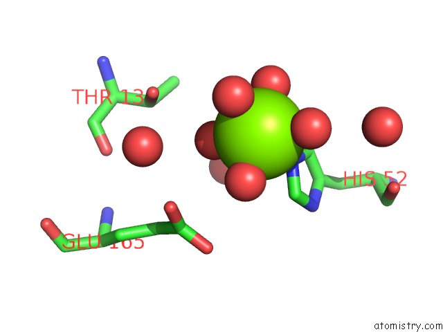
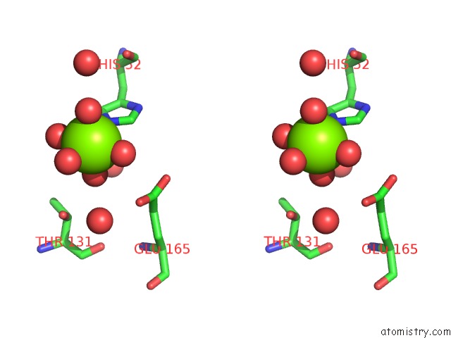
Magnesium binding site 7 out of 7 in 3jr2
Go back to
Magnesium binding site 7 out
of 7 in the X-Ray Crystal Structure of the Mg-Bound 3-Keto-L-Gulonate-6-Phosphate Decarboxylase From Vibrio Cholerae O1 Biovar El Tor Str. N16961
Mono view
Stereo pair view
Mono view
Stereo pair view
A full contact list of Magnesium with other atoms in the Mg binding
site number 7 of X-Ray Crystal Structure of the Mg-Bound 3-Keto-L-Gulonate-6-Phosphate Decarboxylase From Vibrio Cholerae O1 Biovar El Tor Str. N16961 within 5.0Å range:
|
Reference:
B.Nocek,
N.Maltseva,
J.Stam,
W.Anderson,
A.Joachimiak,
Csgid.
Crystal Structure of the Mg-Bound 3-Keto-L-Gulonate-6-Phosphate Decarboxylase From Vibrio Cholerae O1 Biovar El Tor Str. N16961 To Be Published.
Page generated: Sun Aug 10 23:18:48 2025
Last articles
Mg in 6HW8Mg in 6HW7
Mg in 6HW5
Mg in 6HW6
Mg in 6HW4
Mg in 6HVY
Mg in 6HW3
Mg in 6HW0
Mg in 6HW1
Mg in 6HVX