Magnesium »
PDB 3jay-3jwr »
3juk »
Magnesium in PDB 3juk: The Crystal Structure of Udp-Glucose Pyrophosphorylase Complexed with Udp-Glucose
Enzymatic activity of The Crystal Structure of Udp-Glucose Pyrophosphorylase Complexed with Udp-Glucose
All present enzymatic activity of The Crystal Structure of Udp-Glucose Pyrophosphorylase Complexed with Udp-Glucose:
2.7.7.9;
2.7.7.9;
Protein crystallography data
The structure of The Crystal Structure of Udp-Glucose Pyrophosphorylase Complexed with Udp-Glucose, PDB code: 3juk
was solved by
H.Kim,
K.K.Kim,
with X-Ray Crystallography technique. A brief refinement statistics is given in the table below:
| Resolution Low / High (Å) | 50.00 / 2.30 |
| Space group | C 1 2 1 |
| Cell size a, b, c (Å), α, β, γ (°) | 101.444, 74.393, 167.123, 90.00, 97.91, 90.00 |
| R / Rfree (%) | 23 / 27.6 |
Magnesium Binding Sites:
Pages:
>>> Page 1 <<< Page 2, Binding sites: 11 - 12;Binding sites:
The binding sites of Magnesium atom in the The Crystal Structure of Udp-Glucose Pyrophosphorylase Complexed with Udp-Glucose (pdb code 3juk). This binding sites where shown within 5.0 Angstroms radius around Magnesium atom.In total 12 binding sites of Magnesium where determined in the The Crystal Structure of Udp-Glucose Pyrophosphorylase Complexed with Udp-Glucose, PDB code: 3juk:
Jump to Magnesium binding site number: 1; 2; 3; 4; 5; 6; 7; 8; 9; 10;
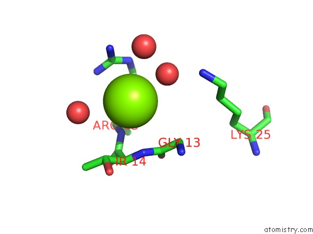
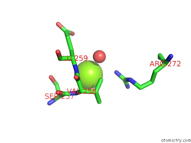
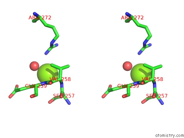
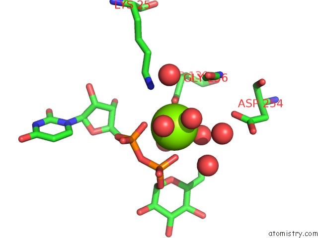
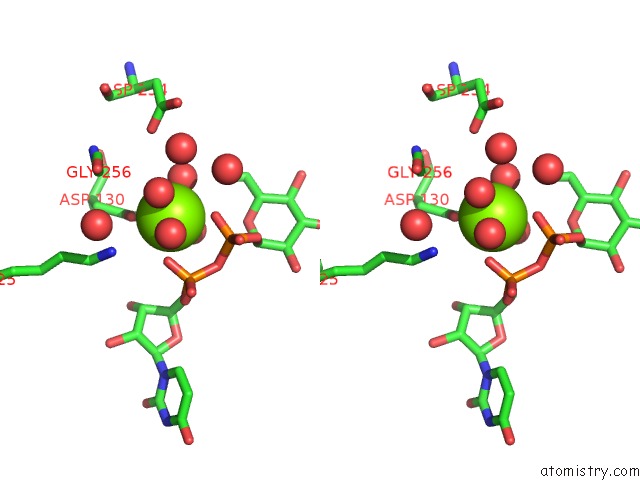
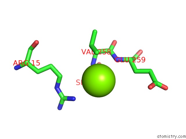
Magnesium binding site 1 out of 12 in 3juk
Go back to
Magnesium binding site 1 out
of 12 in the The Crystal Structure of Udp-Glucose Pyrophosphorylase Complexed with Udp-Glucose
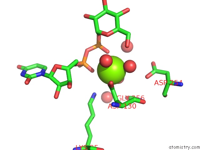
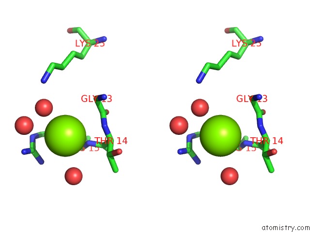
Mono view
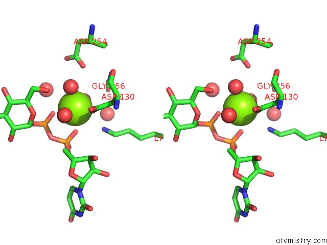
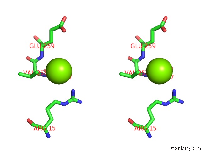
Stereo pair view
Mono view
Stereo pair view
A full contact list of Magnesium with other atoms in the Mg binding
site number 1 of The Crystal Structure of Udp-Glucose Pyrophosphorylase Complexed with Udp-Glucose within 5.0Å range:
|
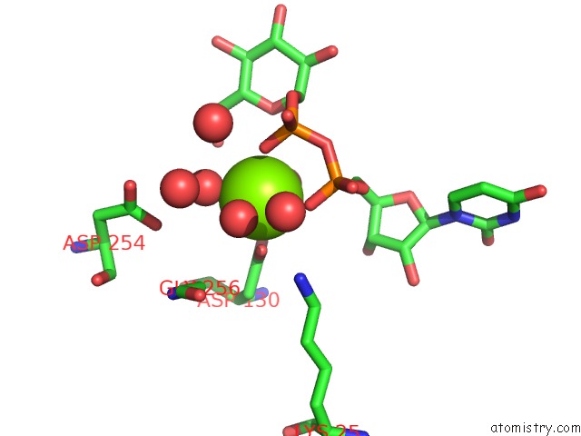
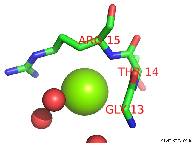
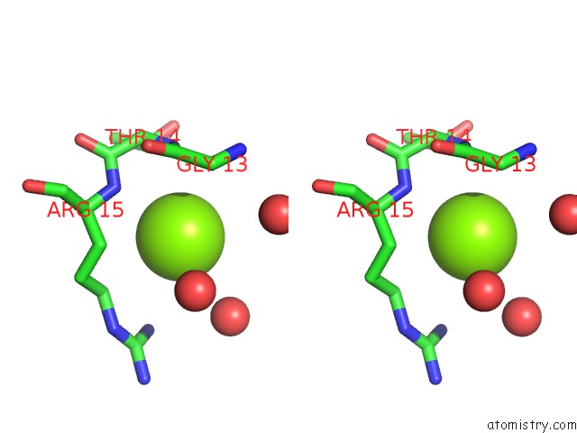
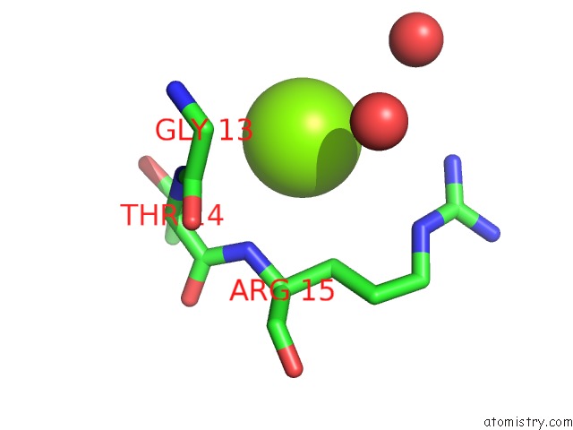
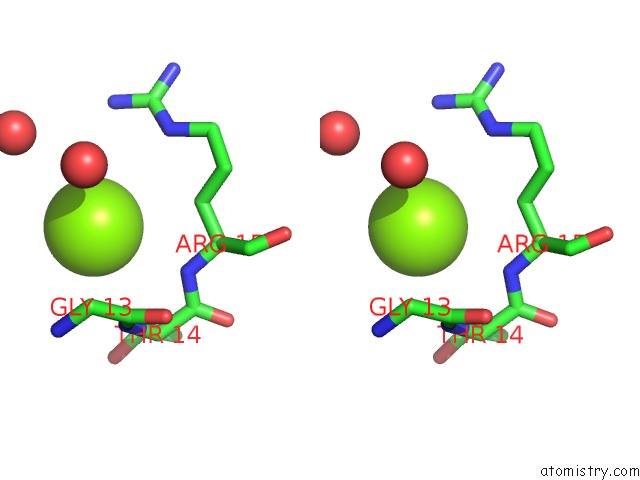
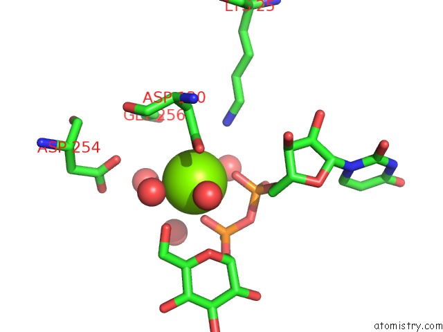
Magnesium binding site 2 out of 12 in 3juk
Go back to
Magnesium binding site 2 out
of 12 in the The Crystal Structure of Udp-Glucose Pyrophosphorylase Complexed with Udp-Glucose
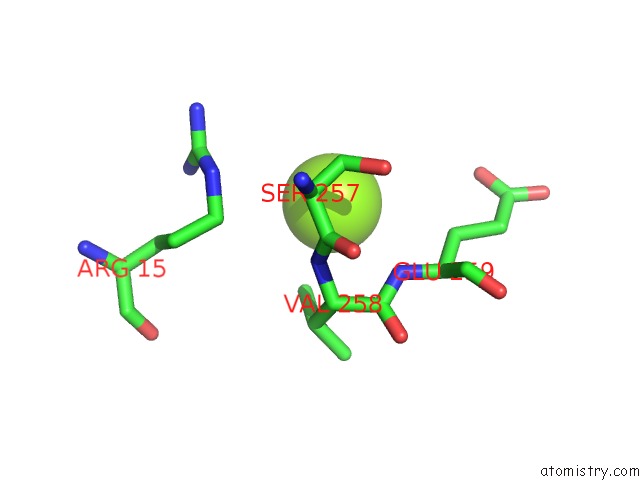
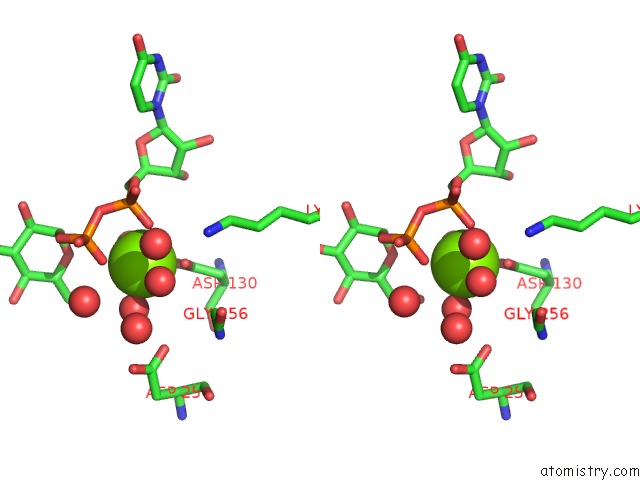
Mono view
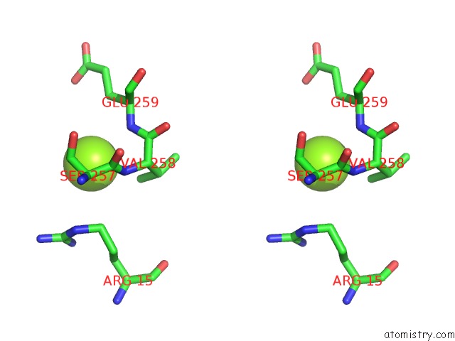
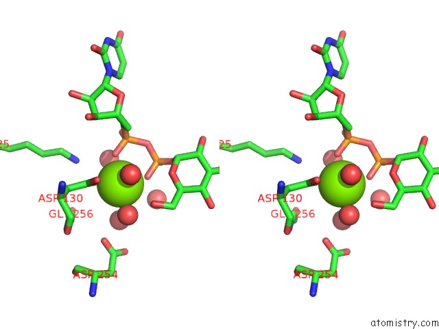
Stereo pair view
Mono view
Stereo pair view
A full contact list of Magnesium with other atoms in the Mg binding
site number 2 of The Crystal Structure of Udp-Glucose Pyrophosphorylase Complexed with Udp-Glucose within 5.0Å range:
|
Magnesium binding site 3 out of 12 in 3juk
Go back to
Magnesium binding site 3 out
of 12 in the The Crystal Structure of Udp-Glucose Pyrophosphorylase Complexed with Udp-Glucose
Mono view
Stereo pair view
Mono view
Stereo pair view
A full contact list of Magnesium with other atoms in the Mg binding
site number 3 of The Crystal Structure of Udp-Glucose Pyrophosphorylase Complexed with Udp-Glucose within 5.0Å range:
|
Magnesium binding site 4 out of 12 in 3juk
Go back to
Magnesium binding site 4 out
of 12 in the The Crystal Structure of Udp-Glucose Pyrophosphorylase Complexed with Udp-Glucose
Mono view
Stereo pair view
Mono view
Stereo pair view
A full contact list of Magnesium with other atoms in the Mg binding
site number 4 of The Crystal Structure of Udp-Glucose Pyrophosphorylase Complexed with Udp-Glucose within 5.0Å range:
|
Magnesium binding site 5 out of 12 in 3juk
Go back to
Magnesium binding site 5 out
of 12 in the The Crystal Structure of Udp-Glucose Pyrophosphorylase Complexed with Udp-Glucose
Mono view
Stereo pair view
Mono view
Stereo pair view
A full contact list of Magnesium with other atoms in the Mg binding
site number 5 of The Crystal Structure of Udp-Glucose Pyrophosphorylase Complexed with Udp-Glucose within 5.0Å range:
|
Magnesium binding site 6 out of 12 in 3juk
Go back to
Magnesium binding site 6 out
of 12 in the The Crystal Structure of Udp-Glucose Pyrophosphorylase Complexed with Udp-Glucose
Mono view
Stereo pair view
Mono view
Stereo pair view
A full contact list of Magnesium with other atoms in the Mg binding
site number 6 of The Crystal Structure of Udp-Glucose Pyrophosphorylase Complexed with Udp-Glucose within 5.0Å range:
|
Magnesium binding site 7 out of 12 in 3juk
Go back to
Magnesium binding site 7 out
of 12 in the The Crystal Structure of Udp-Glucose Pyrophosphorylase Complexed with Udp-Glucose
Mono view
Stereo pair view
Mono view
Stereo pair view
A full contact list of Magnesium with other atoms in the Mg binding
site number 7 of The Crystal Structure of Udp-Glucose Pyrophosphorylase Complexed with Udp-Glucose within 5.0Å range:
|
Magnesium binding site 8 out of 12 in 3juk
Go back to
Magnesium binding site 8 out
of 12 in the The Crystal Structure of Udp-Glucose Pyrophosphorylase Complexed with Udp-Glucose
Mono view
Stereo pair view
Mono view
Stereo pair view
A full contact list of Magnesium with other atoms in the Mg binding
site number 8 of The Crystal Structure of Udp-Glucose Pyrophosphorylase Complexed with Udp-Glucose within 5.0Å range:
|
Magnesium binding site 9 out of 12 in 3juk
Go back to
Magnesium binding site 9 out
of 12 in the The Crystal Structure of Udp-Glucose Pyrophosphorylase Complexed with Udp-Glucose
Mono view
Stereo pair view
Mono view
Stereo pair view
A full contact list of Magnesium with other atoms in the Mg binding
site number 9 of The Crystal Structure of Udp-Glucose Pyrophosphorylase Complexed with Udp-Glucose within 5.0Å range:
|
Magnesium binding site 10 out of 12 in 3juk
Go back to
Magnesium binding site 10 out
of 12 in the The Crystal Structure of Udp-Glucose Pyrophosphorylase Complexed with Udp-Glucose
Mono view
Stereo pair view
Mono view
Stereo pair view
A full contact list of Magnesium with other atoms in the Mg binding
site number 10 of The Crystal Structure of Udp-Glucose Pyrophosphorylase Complexed with Udp-Glucose within 5.0Å range:
|
Reference:
H.Kim,
J.Choi,
T.Kim,
N.K.Lokanath,
S.C.Ha,
S.W.Suh,
H.-Y.Hwang,
K.K.Kim.
Structural Basis For the Reaction Mechanism of Udp-Glucose Pyrophosphorylase Mol.Cells V. 29 397 2010.
ISSN: ISSN 1016-8478
PubMed: 20238176
DOI: 10.1007/S10059-010-0047-6
Page generated: Wed Aug 14 17:14:08 2024
ISSN: ISSN 1016-8478
PubMed: 20238176
DOI: 10.1007/S10059-010-0047-6
Last articles
Ca in 5UE1Ca in 5UE2
Ca in 5UCQ
Ca in 5UDL
Ca in 5UDK
Ca in 5UD6
Ca in 5UDJ
Ca in 5UDI
Ca in 5UD0
Ca in 5UBJ