Magnesium in PDB 3jw7: Crystal Structure of Dipeptide Epimerase From Enterococcus Faecalis V583 Complexed with Mg and Dipeptide L-Ile-L-Tyr
Protein crystallography data
The structure of Crystal Structure of Dipeptide Epimerase From Enterococcus Faecalis V583 Complexed with Mg and Dipeptide L-Ile-L-Tyr, PDB code: 3jw7
was solved by
A.A.Fedorov,
E.V.Fedorov,
H.J.Imker,
A.Sakai,
J.A.Gerlt,
S.C.Almo,
with X-Ray Crystallography technique. A brief refinement statistics is given in the table below:
Resolution Low / High (Å)
|
25.00 /
1.80
|
Space group
|
C 1 2 1
|
Cell size a, b, c (Å), α, β, γ (°)
|
194.918,
187.442,
91.894,
90.00,
89.99,
90.00
|
R / Rfree (%)
|
22.1 /
25.4
|
Magnesium Binding Sites:
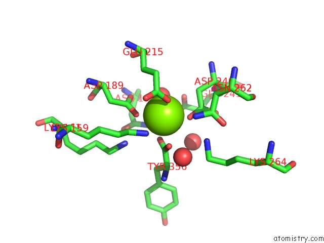
The binding sites of Magnesium atom in the Crystal Structure of Dipeptide Epimerase From Enterococcus Faecalis V583 Complexed with Mg and Dipeptide L-Ile-L-Tyr
(pdb code 3jw7). This binding sites where shown within
5.0 Angstroms radius around Magnesium atom.
In total 8 binding sites of Magnesium where determined in the
Crystal Structure of Dipeptide Epimerase From Enterococcus Faecalis V583 Complexed with Mg and Dipeptide L-Ile-L-Tyr, PDB code: 3jw7:
Jump to Magnesium binding site number:
1;
2;
3;
4;
5;
6;
7;
8;
Magnesium binding site 1 out
of 8 in 3jw7
Go back to
Magnesium Binding Sites List in 3jw7
Magnesium binding site 1 out
of 8 in the Crystal Structure of Dipeptide Epimerase From Enterococcus Faecalis V583 Complexed with Mg and Dipeptide L-Ile-L-Tyr
Mono view
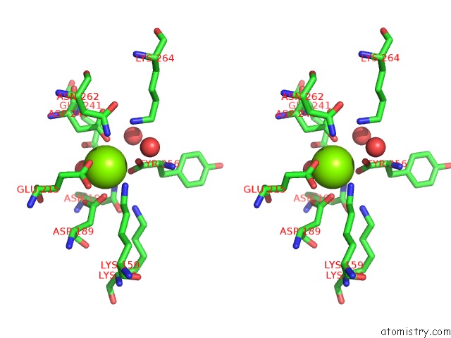
Stereo pair view
|
A full contact list of Magnesium with other atoms in the Mg binding
site number 1 of Crystal Structure of Dipeptide Epimerase From Enterococcus Faecalis V583 Complexed with Mg and Dipeptide L-Ile-L-Tyr within 5.0Å range:
probe
|
atom
|
residue
|
distance (Å)
|
B
|
Occ
|
A:Mg358
b:18.9
occ:1.00
|
OD2
|
A:ASP189
|
2.3
|
15.3
|
1.0
|
OE2
|
A:GLU215
|
2.3
|
19.3
|
1.0
|
OXT
|
A:TYR356
|
2.3
|
16.0
|
1.0
|
OD2
|
A:ASP240
|
2.3
|
17.5
|
1.0
|
O
|
A:HOH360
|
2.4
|
14.2
|
1.0
|
O
|
A:TYR356
|
2.8
|
16.3
|
1.0
|
C
|
A:TYR356
|
2.9
|
18.0
|
1.0
|
CD
|
A:GLU215
|
3.2
|
21.8
|
1.0
|
CG
|
A:ASP189
|
3.2
|
16.3
|
1.0
|
CG
|
A:ASP240
|
3.3
|
18.2
|
1.0
|
OD1
|
A:ASP189
|
3.5
|
15.5
|
1.0
|
NZ
|
A:LYS159
|
3.6
|
16.5
|
1.0
|
CB
|
A:ASP240
|
3.7
|
16.1
|
1.0
|
OE1
|
A:GLU215
|
3.8
|
19.5
|
1.0
|
ND2
|
A:ASN191
|
4.0
|
16.0
|
1.0
|
NZ
|
A:LYS161
|
4.1
|
13.1
|
1.0
|
ND2
|
A:ASN262
|
4.1
|
19.0
|
1.0
|
CG
|
A:GLU215
|
4.1
|
22.2
|
1.0
|
O
|
A:HOH648
|
4.2
|
28.8
|
1.0
|
NZ
|
A:LYS264
|
4.2
|
18.8
|
1.0
|
CB
|
A:GLU215
|
4.3
|
20.7
|
1.0
|
CE
|
A:LYS159
|
4.3
|
17.8
|
1.0
|
CA
|
A:TYR356
|
4.4
|
18.3
|
1.0
|
OD1
|
A:ASP240
|
4.4
|
16.6
|
1.0
|
OE1
|
A:GLU241
|
4.4
|
17.4
|
1.0
|
CB
|
A:ASP189
|
4.6
|
14.2
|
1.0
|
CG
|
A:ASN191
|
4.8
|
18.1
|
1.0
|
O
|
A:HOH369
|
4.8
|
15.5
|
1.0
|
OD1
|
A:ASN191
|
5.0
|
17.1
|
1.0
|
|
Magnesium binding site 2 out
of 8 in 3jw7
Go back to
Magnesium Binding Sites List in 3jw7
Magnesium binding site 2 out
of 8 in the Crystal Structure of Dipeptide Epimerase From Enterococcus Faecalis V583 Complexed with Mg and Dipeptide L-Ile-L-Tyr
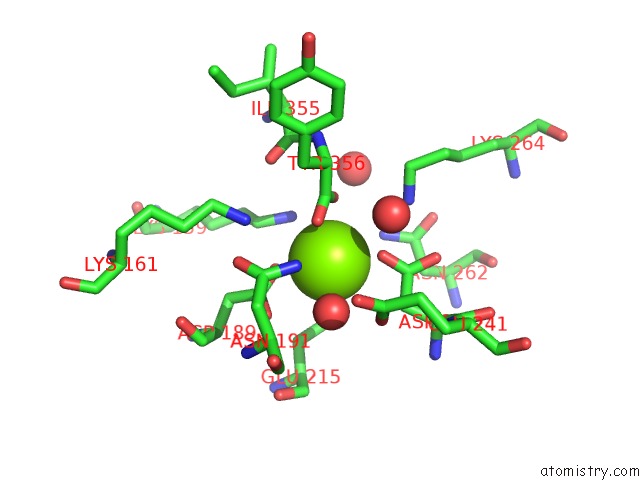
Mono view
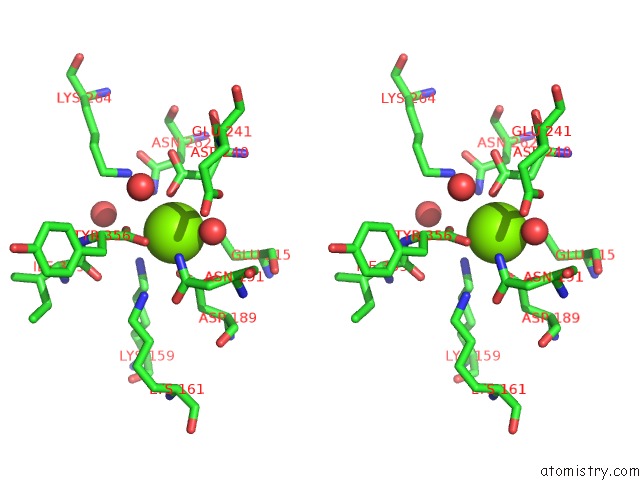
Stereo pair view
|
A full contact list of Magnesium with other atoms in the Mg binding
site number 2 of Crystal Structure of Dipeptide Epimerase From Enterococcus Faecalis V583 Complexed with Mg and Dipeptide L-Ile-L-Tyr within 5.0Å range:
probe
|
atom
|
residue
|
distance (Å)
|
B
|
Occ
|
B:Mg358
b:18.7
occ:1.00
|
OE2
|
B:GLU215
|
2.3
|
21.6
|
1.0
|
OD2
|
B:ASP189
|
2.3
|
13.5
|
1.0
|
OXT
|
B:TYR356
|
2.3
|
15.7
|
1.0
|
OD2
|
B:ASP240
|
2.3
|
17.1
|
1.0
|
O
|
B:HOH359
|
2.4
|
14.9
|
1.0
|
O
|
B:TYR356
|
2.7
|
15.1
|
1.0
|
C
|
B:TYR356
|
2.8
|
15.7
|
1.0
|
CD
|
B:GLU215
|
3.2
|
23.2
|
1.0
|
CG
|
B:ASP189
|
3.2
|
17.0
|
1.0
|
CG
|
B:ASP240
|
3.3
|
17.9
|
1.0
|
OD1
|
B:ASP189
|
3.5
|
14.4
|
1.0
|
NZ
|
B:LYS159
|
3.6
|
17.0
|
1.0
|
CB
|
B:ASP240
|
3.7
|
14.7
|
1.0
|
OE1
|
B:GLU215
|
3.9
|
21.1
|
1.0
|
ND2
|
B:ASN191
|
4.0
|
15.7
|
1.0
|
CG
|
B:GLU215
|
4.0
|
23.4
|
1.0
|
NZ
|
B:LYS161
|
4.0
|
13.2
|
1.0
|
O
|
B:HOH1322
|
4.2
|
28.6
|
1.0
|
ND2
|
B:ASN262
|
4.2
|
19.5
|
1.0
|
NZ
|
B:LYS264
|
4.3
|
18.9
|
1.0
|
CE
|
B:LYS159
|
4.3
|
16.9
|
1.0
|
CA
|
B:TYR356
|
4.3
|
16.5
|
1.0
|
OE1
|
B:GLU241
|
4.3
|
16.8
|
1.0
|
CB
|
B:GLU215
|
4.4
|
21.1
|
1.0
|
OD1
|
B:ASP240
|
4.4
|
16.4
|
1.0
|
CB
|
B:ASP189
|
4.6
|
13.7
|
1.0
|
O
|
B:HOH369
|
4.8
|
15.6
|
1.0
|
CG
|
B:ASN191
|
4.8
|
16.8
|
1.0
|
O
|
B:ILE355
|
4.9
|
18.6
|
1.0
|
OD1
|
B:ASN191
|
4.9
|
18.0
|
1.0
|
CB
|
B:TYR356
|
5.0
|
17.1
|
1.0
|
CD
|
B:GLU241
|
5.0
|
19.1
|
1.0
|
|
Magnesium binding site 3 out
of 8 in 3jw7
Go back to
Magnesium Binding Sites List in 3jw7
Magnesium binding site 3 out
of 8 in the Crystal Structure of Dipeptide Epimerase From Enterococcus Faecalis V583 Complexed with Mg and Dipeptide L-Ile-L-Tyr
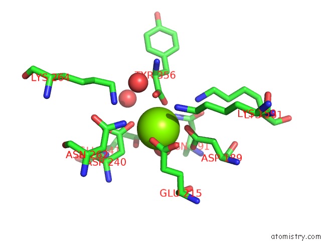
Mono view
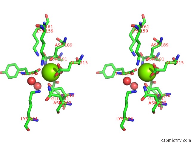
Stereo pair view
|
A full contact list of Magnesium with other atoms in the Mg binding
site number 3 of Crystal Structure of Dipeptide Epimerase From Enterococcus Faecalis V583 Complexed with Mg and Dipeptide L-Ile-L-Tyr within 5.0Å range:
probe
|
atom
|
residue
|
distance (Å)
|
B
|
Occ
|
C:Mg358
b:30.0
occ:1.00
|
OE2
|
C:GLU215
|
2.2
|
29.0
|
1.0
|
OD2
|
C:ASP240
|
2.3
|
28.4
|
1.0
|
OD2
|
C:ASP189
|
2.3
|
24.7
|
1.0
|
O
|
C:HOH363
|
2.4
|
30.8
|
1.0
|
OXT
|
C:TYR356
|
2.5
|
29.2
|
1.0
|
O
|
C:TYR356
|
2.8
|
28.7
|
1.0
|
C
|
C:TYR356
|
3.0
|
30.2
|
1.0
|
CD
|
C:GLU215
|
3.0
|
28.9
|
1.0
|
CG
|
C:ASP189
|
3.2
|
25.4
|
1.0
|
CG
|
C:ASP240
|
3.3
|
26.2
|
1.0
|
OD1
|
C:ASP189
|
3.5
|
25.0
|
1.0
|
NZ
|
C:LYS159
|
3.5
|
28.9
|
1.0
|
CB
|
C:ASP240
|
3.6
|
24.5
|
1.0
|
OE1
|
C:GLU215
|
3.7
|
31.7
|
1.0
|
CG
|
C:GLU215
|
3.8
|
29.7
|
1.0
|
NZ
|
C:LYS161
|
4.1
|
24.5
|
1.0
|
ND2
|
C:ASN191
|
4.1
|
24.7
|
1.0
|
ND2
|
C:ASN262
|
4.1
|
26.7
|
1.0
|
NZ
|
C:LYS264
|
4.2
|
26.7
|
1.0
|
CB
|
C:GLU215
|
4.2
|
28.3
|
1.0
|
OE1
|
C:GLU241
|
4.3
|
23.6
|
1.0
|
CE
|
C:LYS159
|
4.3
|
29.7
|
1.0
|
O
|
C:HOH1430
|
4.3
|
30.3
|
1.0
|
OD1
|
C:ASP240
|
4.4
|
24.5
|
1.0
|
CA
|
C:TYR356
|
4.5
|
30.3
|
1.0
|
CB
|
C:ASP189
|
4.5
|
25.6
|
1.0
|
O
|
C:HOH364
|
4.9
|
26.1
|
1.0
|
CG
|
C:ASN191
|
4.9
|
26.3
|
1.0
|
|
Magnesium binding site 4 out
of 8 in 3jw7
Go back to
Magnesium Binding Sites List in 3jw7
Magnesium binding site 4 out
of 8 in the Crystal Structure of Dipeptide Epimerase From Enterococcus Faecalis V583 Complexed with Mg and Dipeptide L-Ile-L-Tyr
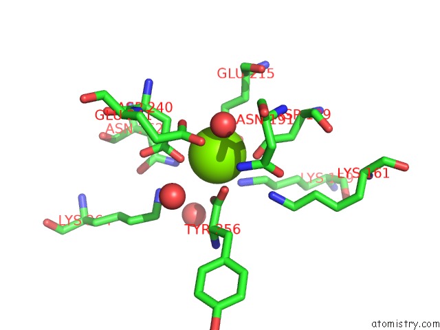
Mono view
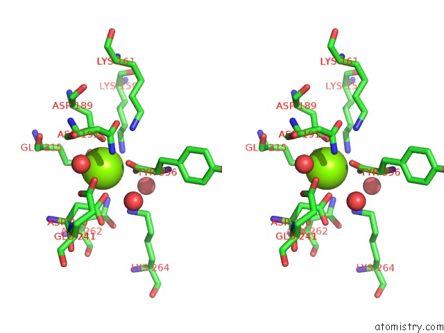
Stereo pair view
|
A full contact list of Magnesium with other atoms in the Mg binding
site number 4 of Crystal Structure of Dipeptide Epimerase From Enterococcus Faecalis V583 Complexed with Mg and Dipeptide L-Ile-L-Tyr within 5.0Å range:
probe
|
atom
|
residue
|
distance (Å)
|
B
|
Occ
|
D:Mg358
b:29.4
occ:1.00
|
OE2
|
D:GLU215
|
2.1
|
30.1
|
1.0
|
OD2
|
D:ASP240
|
2.3
|
27.0
|
1.0
|
OD2
|
D:ASP189
|
2.4
|
25.5
|
1.0
|
O
|
D:HOH925
|
2.4
|
30.9
|
1.0
|
OXT
|
D:TYR356
|
2.5
|
29.9
|
1.0
|
O
|
D:TYR356
|
2.8
|
30.8
|
1.0
|
CD
|
D:GLU215
|
2.9
|
29.7
|
1.0
|
C
|
D:TYR356
|
3.0
|
32.1
|
1.0
|
CG
|
D:ASP189
|
3.3
|
26.2
|
1.0
|
CG
|
D:ASP240
|
3.3
|
25.3
|
1.0
|
NZ
|
D:LYS159
|
3.5
|
27.7
|
1.0
|
OD1
|
D:ASP189
|
3.6
|
25.4
|
1.0
|
OE1
|
D:GLU215
|
3.6
|
31.5
|
1.0
|
CB
|
D:ASP240
|
3.6
|
23.7
|
1.0
|
CG
|
D:GLU215
|
3.8
|
29.4
|
1.0
|
ND2
|
D:ASN262
|
4.1
|
28.6
|
1.0
|
NZ
|
D:LYS161
|
4.1
|
22.9
|
1.0
|
ND2
|
D:ASN191
|
4.1
|
25.6
|
1.0
|
CB
|
D:GLU215
|
4.2
|
27.8
|
1.0
|
CE
|
D:LYS159
|
4.2
|
27.4
|
1.0
|
NZ
|
D:LYS264
|
4.3
|
29.0
|
1.0
|
O
|
D:HOH1007
|
4.4
|
29.9
|
1.0
|
OD1
|
D:ASP240
|
4.4
|
25.3
|
1.0
|
OE1
|
D:GLU241
|
4.4
|
22.7
|
1.0
|
CA
|
D:TYR356
|
4.5
|
31.8
|
1.0
|
CB
|
D:ASP189
|
4.6
|
26.3
|
1.0
|
CG
|
D:ASN191
|
4.9
|
25.3
|
1.0
|
O
|
D:HOH397
|
4.9
|
28.2
|
1.0
|
OD1
|
D:ASN191
|
5.0
|
27.1
|
1.0
|
CG
|
D:ASN262
|
5.0
|
28.6
|
1.0
|
|
Magnesium binding site 5 out
of 8 in 3jw7
Go back to
Magnesium Binding Sites List in 3jw7
Magnesium binding site 5 out
of 8 in the Crystal Structure of Dipeptide Epimerase From Enterococcus Faecalis V583 Complexed with Mg and Dipeptide L-Ile-L-Tyr
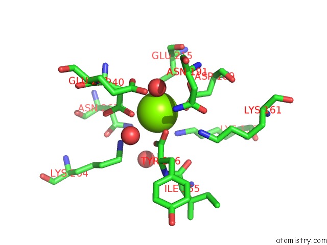
Mono view
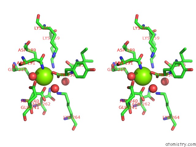
Stereo pair view
|
A full contact list of Magnesium with other atoms in the Mg binding
site number 5 of Crystal Structure of Dipeptide Epimerase From Enterococcus Faecalis V583 Complexed with Mg and Dipeptide L-Ile-L-Tyr within 5.0Å range:
probe
|
atom
|
residue
|
distance (Å)
|
B
|
Occ
|
E:Mg358
b:19.2
occ:1.00
|
OE2
|
E:GLU215
|
2.2
|
21.5
|
1.0
|
OD2
|
E:ASP240
|
2.3
|
19.2
|
1.0
|
O
|
E:HOH368
|
2.3
|
15.6
|
1.0
|
OXT
|
E:TYR356
|
2.3
|
17.1
|
1.0
|
OD2
|
E:ASP189
|
2.4
|
17.0
|
1.0
|
O
|
E:TYR356
|
2.7
|
15.9
|
1.0
|
C
|
E:TYR356
|
2.8
|
14.6
|
1.0
|
CD
|
E:GLU215
|
3.1
|
21.1
|
1.0
|
CG
|
E:ASP240
|
3.2
|
19.3
|
1.0
|
CG
|
E:ASP189
|
3.4
|
17.9
|
1.0
|
NZ
|
E:LYS159
|
3.6
|
15.4
|
1.0
|
CB
|
E:ASP240
|
3.6
|
16.3
|
1.0
|
OD1
|
E:ASP189
|
3.6
|
17.5
|
1.0
|
OE1
|
E:GLU215
|
3.9
|
21.6
|
1.0
|
CG
|
E:GLU215
|
3.9
|
20.9
|
1.0
|
ND2
|
E:ASN191
|
3.9
|
15.1
|
1.0
|
O
|
E:HOH1402
|
4.1
|
36.8
|
1.0
|
NZ
|
E:LYS264
|
4.1
|
15.3
|
1.0
|
NZ
|
E:LYS161
|
4.1
|
13.6
|
1.0
|
CA
|
E:TYR356
|
4.3
|
16.3
|
1.0
|
OD1
|
E:ASP240
|
4.3
|
18.6
|
1.0
|
ND2
|
E:ASN262
|
4.3
|
20.3
|
1.0
|
CE
|
E:LYS159
|
4.4
|
16.0
|
1.0
|
OE1
|
E:GLU241
|
4.4
|
17.1
|
1.0
|
CB
|
E:GLU215
|
4.4
|
16.9
|
1.0
|
CB
|
E:ASP189
|
4.7
|
14.5
|
1.0
|
CG
|
E:ASN191
|
4.8
|
14.8
|
1.0
|
O
|
E:HOH387
|
4.8
|
15.5
|
1.0
|
CB
|
E:TYR356
|
4.9
|
16.6
|
1.0
|
O
|
E:ILE355
|
4.9
|
16.6
|
1.0
|
CD
|
E:GLU241
|
5.0
|
18.7
|
1.0
|
OD1
|
E:ASN191
|
5.0
|
14.8
|
1.0
|
|
Magnesium binding site 6 out
of 8 in 3jw7
Go back to
Magnesium Binding Sites List in 3jw7
Magnesium binding site 6 out
of 8 in the Crystal Structure of Dipeptide Epimerase From Enterococcus Faecalis V583 Complexed with Mg and Dipeptide L-Ile-L-Tyr
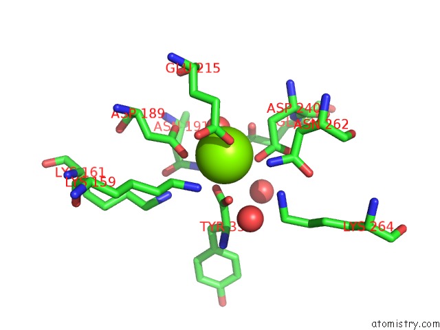
Mono view
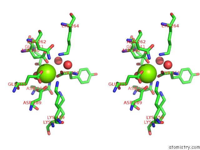
Stereo pair view
|
A full contact list of Magnesium with other atoms in the Mg binding
site number 6 of Crystal Structure of Dipeptide Epimerase From Enterococcus Faecalis V583 Complexed with Mg and Dipeptide L-Ile-L-Tyr within 5.0Å range:
probe
|
atom
|
residue
|
distance (Å)
|
B
|
Occ
|
F:Mg358
b:23.6
occ:1.00
|
OD2
|
F:ASP240
|
2.2
|
25.7
|
1.0
|
OE2
|
F:GLU215
|
2.2
|
24.2
|
1.0
|
O
|
F:HOH393
|
2.2
|
18.4
|
1.0
|
OD2
|
F:ASP189
|
2.4
|
25.1
|
1.0
|
OXT
|
F:TYR356
|
2.4
|
24.5
|
1.0
|
O
|
F:TYR356
|
2.8
|
23.3
|
1.0
|
C
|
F:TYR356
|
2.9
|
22.6
|
1.0
|
CD
|
F:GLU215
|
3.1
|
23.2
|
1.0
|
CG
|
F:ASP240
|
3.1
|
23.8
|
1.0
|
CG
|
F:ASP189
|
3.3
|
23.4
|
1.0
|
CB
|
F:ASP240
|
3.5
|
21.0
|
1.0
|
OD1
|
F:ASP189
|
3.6
|
24.2
|
1.0
|
NZ
|
F:LYS159
|
3.8
|
21.9
|
1.0
|
OE1
|
F:GLU215
|
3.8
|
23.9
|
1.0
|
CG
|
F:GLU215
|
3.9
|
24.4
|
1.0
|
ND2
|
F:ASN191
|
4.1
|
19.9
|
1.0
|
NZ
|
F:LYS264
|
4.1
|
22.8
|
1.0
|
O
|
F:HOH660
|
4.2
|
31.6
|
1.0
|
OD1
|
F:ASP240
|
4.2
|
23.7
|
1.0
|
ND2
|
F:ASN262
|
4.2
|
23.7
|
1.0
|
NZ
|
F:LYS161
|
4.3
|
19.3
|
1.0
|
OE1
|
F:GLU241
|
4.3
|
22.3
|
1.0
|
CB
|
F:GLU215
|
4.3
|
22.8
|
1.0
|
CE
|
F:LYS159
|
4.4
|
24.4
|
1.0
|
CA
|
F:TYR356
|
4.4
|
23.4
|
1.0
|
CB
|
F:ASP189
|
4.7
|
21.0
|
1.0
|
CG
|
F:ASN191
|
4.9
|
22.9
|
1.0
|
O
|
F:HOH390
|
4.9
|
21.1
|
1.0
|
CD
|
F:GLU241
|
4.9
|
23.6
|
1.0
|
|
Magnesium binding site 7 out
of 8 in 3jw7
Go back to
Magnesium Binding Sites List in 3jw7
Magnesium binding site 7 out
of 8 in the Crystal Structure of Dipeptide Epimerase From Enterococcus Faecalis V583 Complexed with Mg and Dipeptide L-Ile-L-Tyr
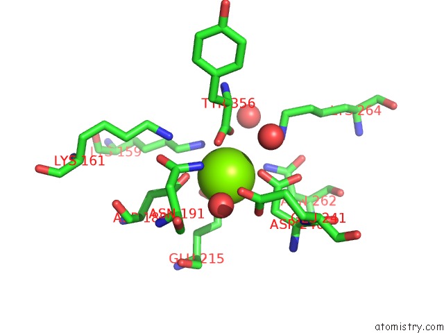
Mono view
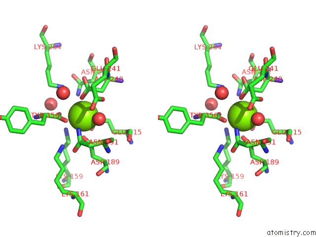
Stereo pair view
|
A full contact list of Magnesium with other atoms in the Mg binding
site number 7 of Crystal Structure of Dipeptide Epimerase From Enterococcus Faecalis V583 Complexed with Mg and Dipeptide L-Ile-L-Tyr within 5.0Å range:
probe
|
atom
|
residue
|
distance (Å)
|
B
|
Occ
|
G:Mg358
b:23.4
occ:1.00
|
OD2
|
G:ASP240
|
2.2
|
22.8
|
1.0
|
OE2
|
G:GLU215
|
2.2
|
24.5
|
1.0
|
O
|
G:HOH384
|
2.3
|
17.7
|
1.0
|
OXT
|
G:TYR356
|
2.4
|
24.0
|
1.0
|
OD2
|
G:ASP189
|
2.4
|
24.5
|
1.0
|
O
|
G:TYR356
|
2.8
|
24.2
|
1.0
|
C
|
G:TYR356
|
2.9
|
23.5
|
1.0
|
CD
|
G:GLU215
|
3.1
|
23.1
|
1.0
|
CG
|
G:ASP240
|
3.2
|
19.7
|
1.0
|
CG
|
G:ASP189
|
3.4
|
22.6
|
1.0
|
CB
|
G:ASP240
|
3.6
|
18.6
|
1.0
|
OD1
|
G:ASP189
|
3.7
|
25.3
|
1.0
|
NZ
|
G:LYS159
|
3.8
|
20.6
|
1.0
|
OE1
|
G:GLU215
|
3.8
|
24.2
|
1.0
|
CG
|
G:GLU215
|
3.9
|
24.0
|
1.0
|
ND2
|
G:ASN191
|
4.0
|
19.0
|
1.0
|
NZ
|
G:LYS264
|
4.1
|
21.8
|
1.0
|
O
|
G:HOH2521
|
4.2
|
26.0
|
1.0
|
NZ
|
G:LYS161
|
4.2
|
18.6
|
1.0
|
ND2
|
G:ASN262
|
4.2
|
22.8
|
1.0
|
OD1
|
G:ASP240
|
4.2
|
21.2
|
1.0
|
OE1
|
G:GLU241
|
4.3
|
23.1
|
1.0
|
CB
|
G:GLU215
|
4.4
|
20.7
|
1.0
|
CE
|
G:LYS159
|
4.4
|
23.4
|
1.0
|
CA
|
G:TYR356
|
4.4
|
23.6
|
1.0
|
CB
|
G:ASP189
|
4.7
|
21.2
|
1.0
|
CG
|
G:ASN191
|
4.8
|
22.7
|
1.0
|
CD
|
G:GLU241
|
4.9
|
22.4
|
1.0
|
O
|
G:HOH406
|
4.9
|
21.9
|
1.0
|
|
Magnesium binding site 8 out
of 8 in 3jw7
Go back to
Magnesium Binding Sites List in 3jw7
Magnesium binding site 8 out
of 8 in the Crystal Structure of Dipeptide Epimerase From Enterococcus Faecalis V583 Complexed with Mg and Dipeptide L-Ile-L-Tyr
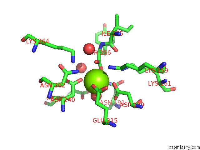
Mono view
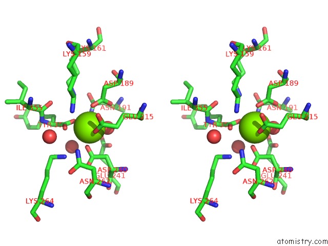
Stereo pair view
|
A full contact list of Magnesium with other atoms in the Mg binding
site number 8 of Crystal Structure of Dipeptide Epimerase From Enterococcus Faecalis V583 Complexed with Mg and Dipeptide L-Ile-L-Tyr within 5.0Å range:
probe
|
atom
|
residue
|
distance (Å)
|
B
|
Occ
|
H:Mg358
b:19.5
occ:1.00
|
OE2
|
H:GLU215
|
2.2
|
21.1
|
1.0
|
OD2
|
H:ASP240
|
2.3
|
16.0
|
1.0
|
O
|
H:HOH359
|
2.3
|
14.7
|
1.0
|
OXT
|
H:TYR356
|
2.3
|
17.0
|
1.0
|
OD2
|
H:ASP189
|
2.4
|
18.2
|
1.0
|
O
|
H:TYR356
|
2.7
|
15.2
|
1.0
|
C
|
H:TYR356
|
2.8
|
16.0
|
1.0
|
CD
|
H:GLU215
|
3.1
|
21.3
|
1.0
|
CG
|
H:ASP240
|
3.2
|
16.7
|
1.0
|
CG
|
H:ASP189
|
3.3
|
18.1
|
1.0
|
OD1
|
H:ASP189
|
3.5
|
17.9
|
1.0
|
NZ
|
H:LYS159
|
3.6
|
15.8
|
1.0
|
CB
|
H:ASP240
|
3.6
|
13.9
|
1.0
|
ND2
|
H:ASN191
|
3.8
|
13.4
|
1.0
|
OE1
|
H:GLU215
|
3.9
|
22.3
|
1.0
|
CG
|
H:GLU215
|
4.0
|
21.7
|
1.0
|
O
|
H:HOH1070
|
4.1
|
31.1
|
1.0
|
NZ
|
H:LYS161
|
4.1
|
11.2
|
1.0
|
NZ
|
H:LYS264
|
4.2
|
16.1
|
1.0
|
CA
|
H:TYR356
|
4.3
|
16.1
|
1.0
|
OD1
|
H:ASP240
|
4.3
|
17.7
|
1.0
|
ND2
|
H:ASN262
|
4.3
|
19.5
|
1.0
|
OE1
|
H:GLU241
|
4.4
|
17.5
|
1.0
|
CE
|
H:LYS159
|
4.4
|
14.9
|
1.0
|
CB
|
H:GLU215
|
4.4
|
17.6
|
1.0
|
CB
|
H:ASP189
|
4.7
|
15.6
|
1.0
|
CG
|
H:ASN191
|
4.7
|
13.5
|
1.0
|
O
|
H:HOH417
|
4.9
|
16.5
|
1.0
|
CB
|
H:TYR356
|
5.0
|
17.5
|
1.0
|
CD
|
H:GLU241
|
5.0
|
18.9
|
1.0
|
O
|
H:ILE355
|
5.0
|
15.2
|
1.0
|
OD1
|
H:ASN191
|
5.0
|
14.6
|
1.0
|
|
Reference:
T.Lukk,
A.Sakai,
C.Kalyanaraman,
S.D.Brown,
H.J.Imker,
L.Song,
A.A.Fedorov,
E.V.Fedorov,
R.Toro,
B.Hillerich,
R.Seidel,
Y.Patskovsky,
M.W.Vetting,
S.K.Nair,
P.C.Babbitt,
S.C.Almo,
J.A.Gerlt,
M.P.Jacobson.
Homology Models Guide Discovery of Diverse Enzyme Specificities Among Dipeptide Epimerases in the Enolase Superfamily. Proc.Natl.Acad.Sci.Usa V. 109 4122 2012.
ISSN: ISSN 0027-8424
PubMed: 22392983
DOI: 10.1073/PNAS.1112081109
Page generated: Wed Aug 14 17:15:12 2024
|