Magnesium »
PDB 3jxq-3k9f »
3k09 »
Magnesium in PDB 3k09: Crystal Structure of the Phosphorylation-Site Mutant S431D of the Kaic Circadian Clock Protein
Enzymatic activity of Crystal Structure of the Phosphorylation-Site Mutant S431D of the Kaic Circadian Clock Protein
All present enzymatic activity of Crystal Structure of the Phosphorylation-Site Mutant S431D of the Kaic Circadian Clock Protein:
2.7.11.1;
2.7.11.1;
Protein crystallography data
The structure of Crystal Structure of the Phosphorylation-Site Mutant S431D of the Kaic Circadian Clock Protein, PDB code: 3k09
was solved by
R.Pattanayek,
M.Egli,
S.Pattanayek,
with X-Ray Crystallography technique. A brief refinement statistics is given in the table below:
| Resolution Low / High (Å) | 30.00 / 3.20 |
| Space group | P 21 21 21 |
| Cell size a, b, c (Å), α, β, γ (°) | 132.500, 135.830, 204.320, 90.00, 90.00, 90.00 |
| R / Rfree (%) | 23.3 / 29.6 |
Magnesium Binding Sites:
Pages:
>>> Page 1 <<< Page 2, Binding sites: 11 - 20;Binding sites:
The binding sites of Magnesium atom in the Crystal Structure of the Phosphorylation-Site Mutant S431D of the Kaic Circadian Clock Protein (pdb code 3k09). This binding sites where shown within 5.0 Angstroms radius around Magnesium atom.In total 20 binding sites of Magnesium where determined in the Crystal Structure of the Phosphorylation-Site Mutant S431D of the Kaic Circadian Clock Protein, PDB code: 3k09:
Jump to Magnesium binding site number: 1; 2; 3; 4; 5; 6; 7; 8; 9; 10;
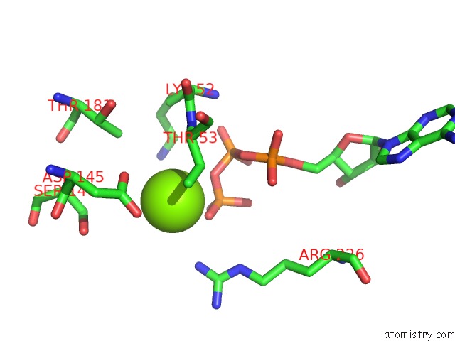
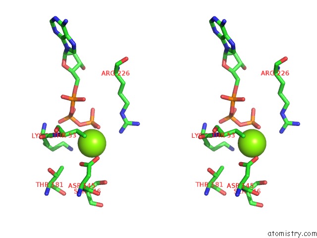
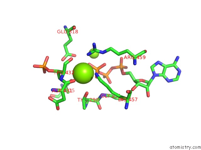
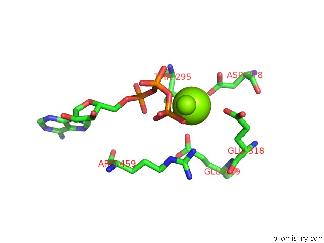
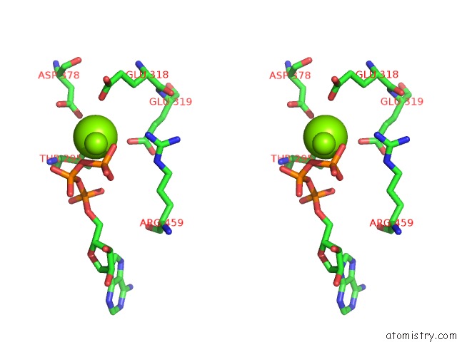
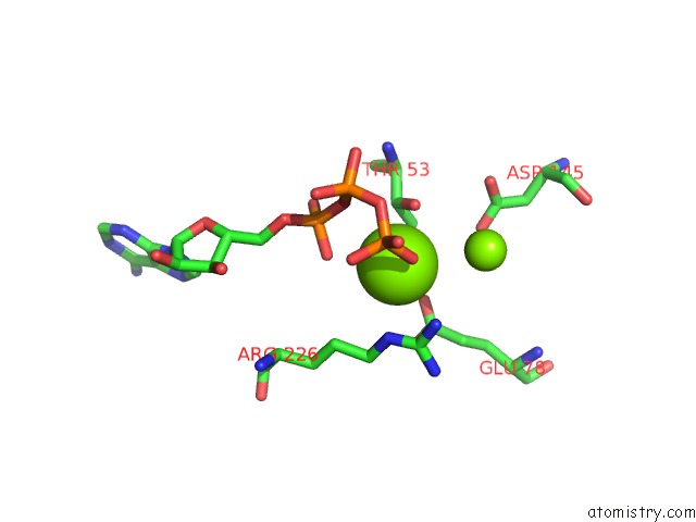
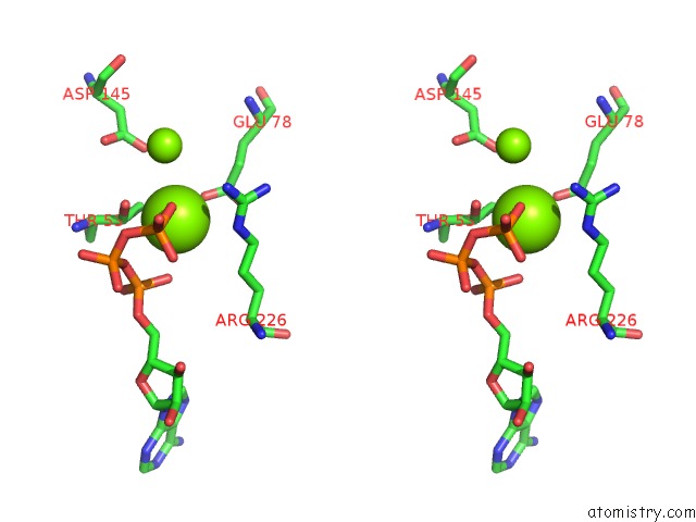
Magnesium binding site 1 out of 20 in 3k09
Go back to
Magnesium binding site 1 out
of 20 in the Crystal Structure of the Phosphorylation-Site Mutant S431D of the Kaic Circadian Clock Protein
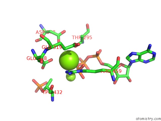
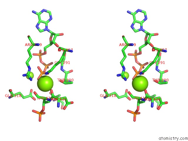
Mono view
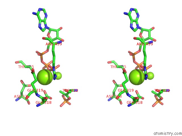
Stereo pair view
Mono view
Stereo pair view
A full contact list of Magnesium with other atoms in the Mg binding
site number 1 of Crystal Structure of the Phosphorylation-Site Mutant S431D of the Kaic Circadian Clock Protein within 5.0Å range:
|
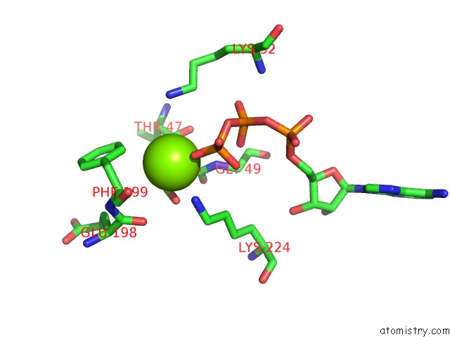
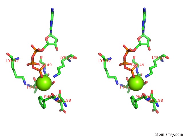
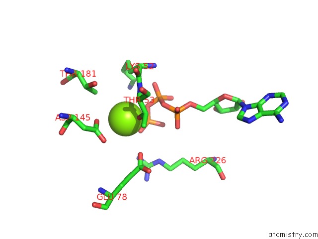
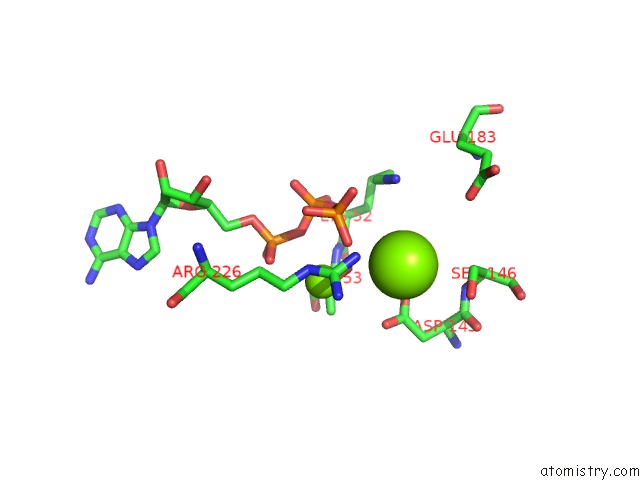
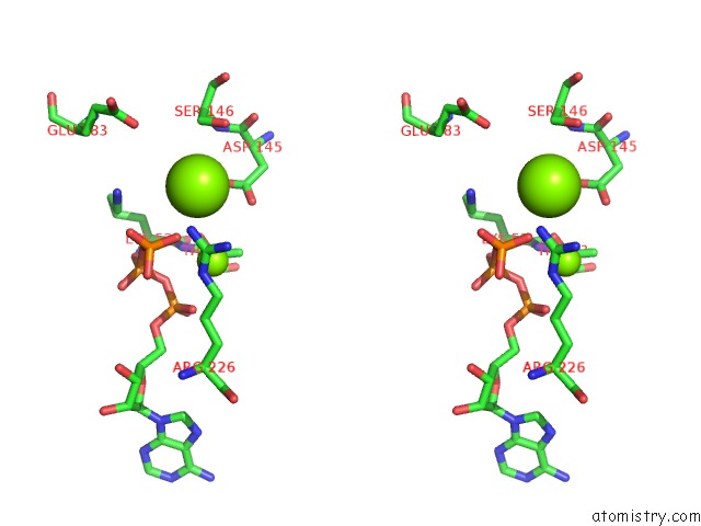
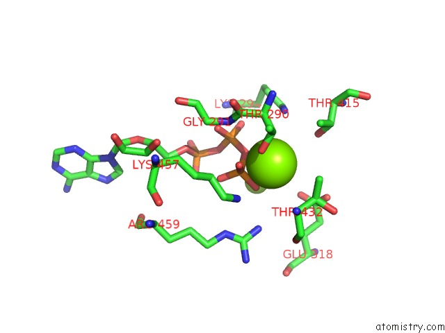
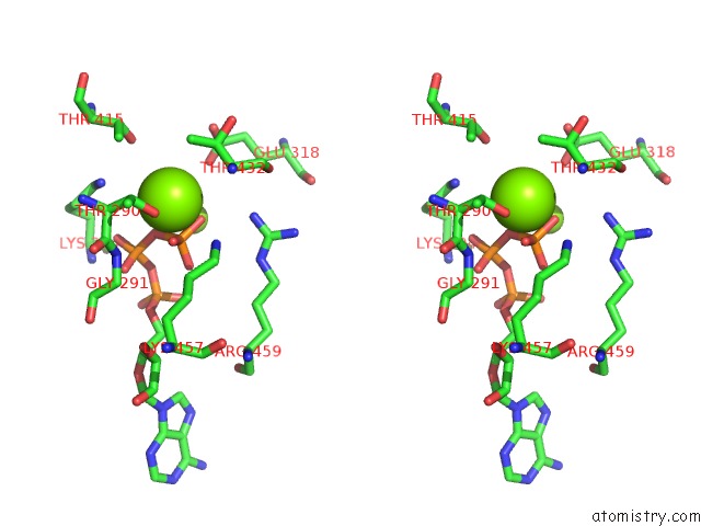
Magnesium binding site 2 out of 20 in 3k09
Go back to
Magnesium binding site 2 out
of 20 in the Crystal Structure of the Phosphorylation-Site Mutant S431D of the Kaic Circadian Clock Protein
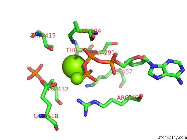
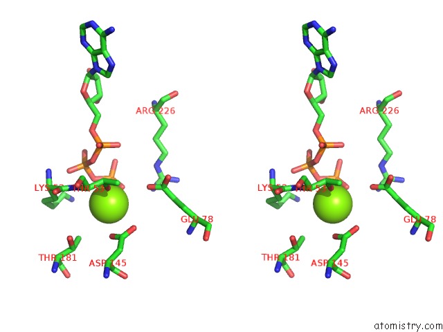
Mono view
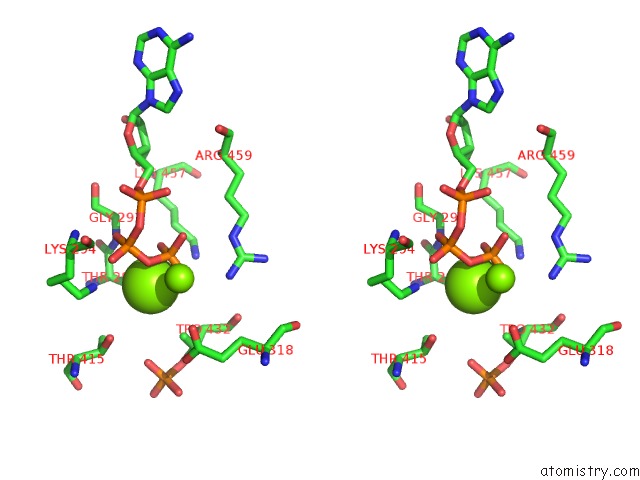
Stereo pair view
Mono view
Stereo pair view
A full contact list of Magnesium with other atoms in the Mg binding
site number 2 of Crystal Structure of the Phosphorylation-Site Mutant S431D of the Kaic Circadian Clock Protein within 5.0Å range:
|
Magnesium binding site 3 out of 20 in 3k09
Go back to
Magnesium binding site 3 out
of 20 in the Crystal Structure of the Phosphorylation-Site Mutant S431D of the Kaic Circadian Clock Protein
Mono view
Stereo pair view
Mono view
Stereo pair view
A full contact list of Magnesium with other atoms in the Mg binding
site number 3 of Crystal Structure of the Phosphorylation-Site Mutant S431D of the Kaic Circadian Clock Protein within 5.0Å range:
|
Magnesium binding site 4 out of 20 in 3k09
Go back to
Magnesium binding site 4 out
of 20 in the Crystal Structure of the Phosphorylation-Site Mutant S431D of the Kaic Circadian Clock Protein
Mono view
Stereo pair view
Mono view
Stereo pair view
A full contact list of Magnesium with other atoms in the Mg binding
site number 4 of Crystal Structure of the Phosphorylation-Site Mutant S431D of the Kaic Circadian Clock Protein within 5.0Å range:
|
Magnesium binding site 5 out of 20 in 3k09
Go back to
Magnesium binding site 5 out
of 20 in the Crystal Structure of the Phosphorylation-Site Mutant S431D of the Kaic Circadian Clock Protein
Mono view
Stereo pair view
Mono view
Stereo pair view
A full contact list of Magnesium with other atoms in the Mg binding
site number 5 of Crystal Structure of the Phosphorylation-Site Mutant S431D of the Kaic Circadian Clock Protein within 5.0Å range:
|
Magnesium binding site 6 out of 20 in 3k09
Go back to
Magnesium binding site 6 out
of 20 in the Crystal Structure of the Phosphorylation-Site Mutant S431D of the Kaic Circadian Clock Protein
Mono view
Stereo pair view
Mono view
Stereo pair view
A full contact list of Magnesium with other atoms in the Mg binding
site number 6 of Crystal Structure of the Phosphorylation-Site Mutant S431D of the Kaic Circadian Clock Protein within 5.0Å range:
|
Magnesium binding site 7 out of 20 in 3k09
Go back to
Magnesium binding site 7 out
of 20 in the Crystal Structure of the Phosphorylation-Site Mutant S431D of the Kaic Circadian Clock Protein
Mono view
Stereo pair view
Mono view
Stereo pair view
A full contact list of Magnesium with other atoms in the Mg binding
site number 7 of Crystal Structure of the Phosphorylation-Site Mutant S431D of the Kaic Circadian Clock Protein within 5.0Å range:
|
Magnesium binding site 8 out of 20 in 3k09
Go back to
Magnesium binding site 8 out
of 20 in the Crystal Structure of the Phosphorylation-Site Mutant S431D of the Kaic Circadian Clock Protein
Mono view
Stereo pair view
Mono view
Stereo pair view
A full contact list of Magnesium with other atoms in the Mg binding
site number 8 of Crystal Structure of the Phosphorylation-Site Mutant S431D of the Kaic Circadian Clock Protein within 5.0Å range:
|
Magnesium binding site 9 out of 20 in 3k09
Go back to
Magnesium binding site 9 out
of 20 in the Crystal Structure of the Phosphorylation-Site Mutant S431D of the Kaic Circadian Clock Protein
Mono view
Stereo pair view
Mono view
Stereo pair view
A full contact list of Magnesium with other atoms in the Mg binding
site number 9 of Crystal Structure of the Phosphorylation-Site Mutant S431D of the Kaic Circadian Clock Protein within 5.0Å range:
|
Magnesium binding site 10 out of 20 in 3k09
Go back to
Magnesium binding site 10 out
of 20 in the Crystal Structure of the Phosphorylation-Site Mutant S431D of the Kaic Circadian Clock Protein
Mono view
Stereo pair view
Mono view
Stereo pair view
A full contact list of Magnesium with other atoms in the Mg binding
site number 10 of Crystal Structure of the Phosphorylation-Site Mutant S431D of the Kaic Circadian Clock Protein within 5.0Å range:
|
Reference:
R.Pattanayek,
T.Mori,
Y.Xu,
S.Pattanayek,
C.H.Johnson,
M.Egli.
Structures of Kaic Circadian Clock Mutant Proteins: A New Phosphorylation Site at T426 and Mechanisms of Kinase, Atpase and Phosphatase. Plos One V. 4 E7529 2009.
ISSN: ESSN 1932-6203
PubMed: 19956664
DOI: 10.1371/JOURNAL.PONE.0007529
Page generated: Wed Aug 14 17:43:40 2024
ISSN: ESSN 1932-6203
PubMed: 19956664
DOI: 10.1371/JOURNAL.PONE.0007529
Last articles
Fe in 2YXOFe in 2YRS
Fe in 2YXC
Fe in 2YNM
Fe in 2YVJ
Fe in 2YP1
Fe in 2YU2
Fe in 2YU1
Fe in 2YQB
Fe in 2YOO