Magnesium »
PDB 3jxq-3k9f »
3k0f »
Magnesium in PDB 3k0f: Crystal Structure of the Phosphorylation-Site Double Mutant T426A/T432A of the Kaic Circadian Clock Protein
Enzymatic activity of Crystal Structure of the Phosphorylation-Site Double Mutant T426A/T432A of the Kaic Circadian Clock Protein
All present enzymatic activity of Crystal Structure of the Phosphorylation-Site Double Mutant T426A/T432A of the Kaic Circadian Clock Protein:
2.7.11.17;
2.7.11.17;
Protein crystallography data
The structure of Crystal Structure of the Phosphorylation-Site Double Mutant T426A/T432A of the Kaic Circadian Clock Protein, PDB code: 3k0f
was solved by
R.Pattanayek,
M.Egli,
S.Pattanayek,
with X-Ray Crystallography technique. A brief refinement statistics is given in the table below:
| Resolution Low / High (Å) | 30.00 / 3.00 |
| Space group | P 21 21 21 |
| Cell size a, b, c (Å), α, β, γ (°) | 132.927, 135.415, 205.618, 90.00, 90.00, 90.00 |
| R / Rfree (%) | 22.9 / 28.8 |
Magnesium Binding Sites:
The binding sites of Magnesium atom in the Crystal Structure of the Phosphorylation-Site Double Mutant T426A/T432A of the Kaic Circadian Clock Protein
(pdb code 3k0f). This binding sites where shown within
5.0 Angstroms radius around Magnesium atom.
In total 9 binding sites of Magnesium where determined in the Crystal Structure of the Phosphorylation-Site Double Mutant T426A/T432A of the Kaic Circadian Clock Protein, PDB code: 3k0f:
Jump to Magnesium binding site number: 1; 2; 3; 4; 5; 6; 7; 8; 9;
In total 9 binding sites of Magnesium where determined in the Crystal Structure of the Phosphorylation-Site Double Mutant T426A/T432A of the Kaic Circadian Clock Protein, PDB code: 3k0f:
Jump to Magnesium binding site number: 1; 2; 3; 4; 5; 6; 7; 8; 9;
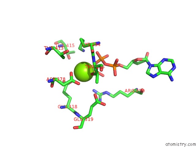
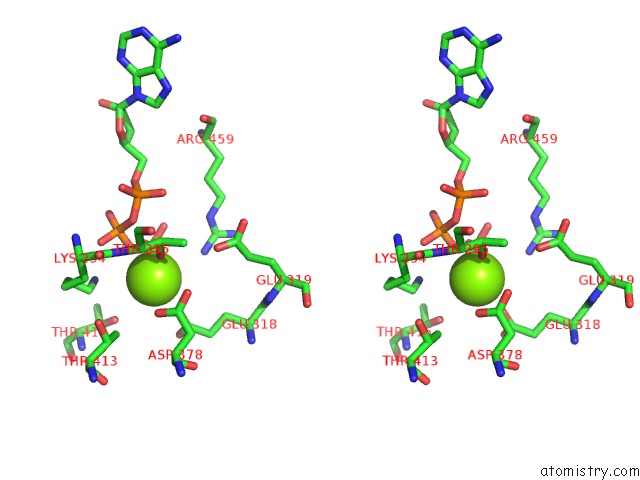
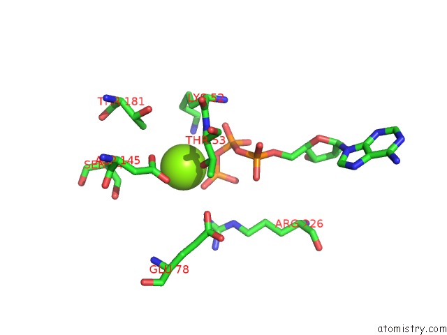
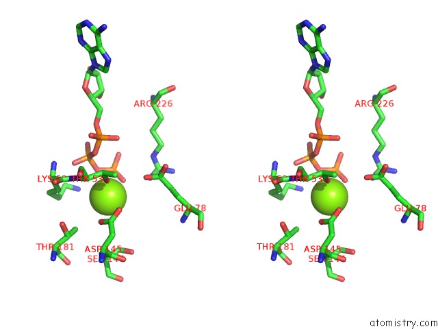
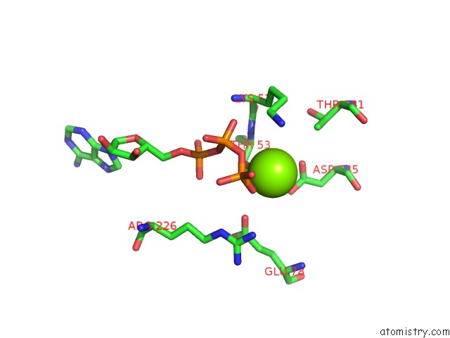
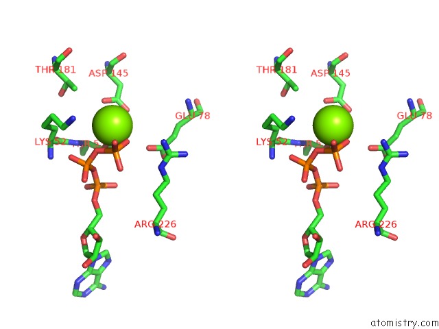
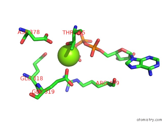
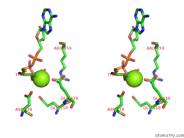
Magnesium binding site 1 out of 9 in 3k0f
Go back to
Magnesium binding site 1 out
of 9 in the Crystal Structure of the Phosphorylation-Site Double Mutant T426A/T432A of the Kaic Circadian Clock Protein
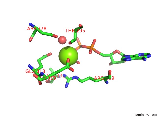
Mono view
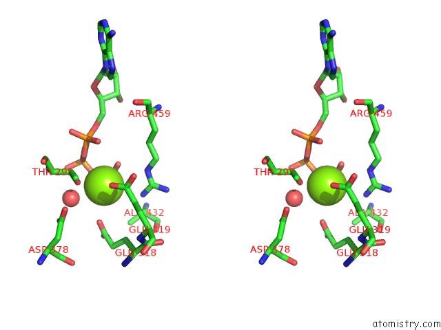
Stereo pair view
Mono view
Stereo pair view
A full contact list of Magnesium with other atoms in the Mg binding
site number 1 of Crystal Structure of the Phosphorylation-Site Double Mutant T426A/T432A of the Kaic Circadian Clock Protein within 5.0Å range:
|
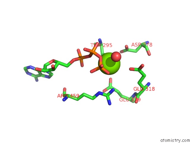
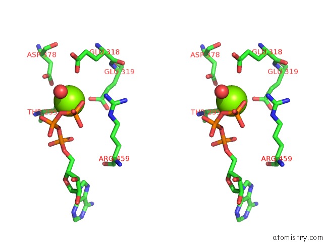
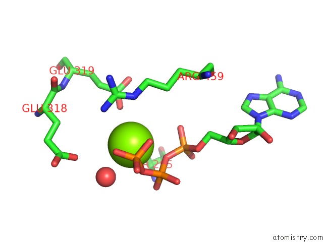
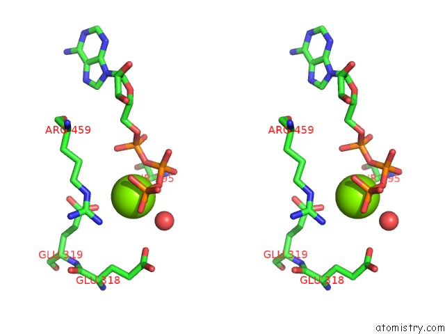
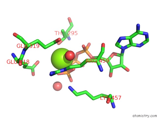
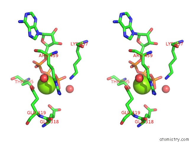
Magnesium binding site 2 out of 9 in 3k0f
Go back to
Magnesium binding site 2 out
of 9 in the Crystal Structure of the Phosphorylation-Site Double Mutant T426A/T432A of the Kaic Circadian Clock Protein
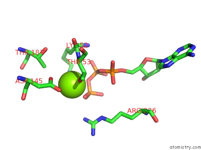
Mono view
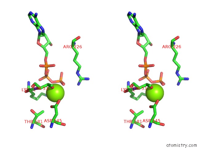
Stereo pair view
Mono view
Stereo pair view
A full contact list of Magnesium with other atoms in the Mg binding
site number 2 of Crystal Structure of the Phosphorylation-Site Double Mutant T426A/T432A of the Kaic Circadian Clock Protein within 5.0Å range:
|
Magnesium binding site 3 out of 9 in 3k0f
Go back to
Magnesium binding site 3 out
of 9 in the Crystal Structure of the Phosphorylation-Site Double Mutant T426A/T432A of the Kaic Circadian Clock Protein
Mono view
Stereo pair view
Mono view
Stereo pair view
A full contact list of Magnesium with other atoms in the Mg binding
site number 3 of Crystal Structure of the Phosphorylation-Site Double Mutant T426A/T432A of the Kaic Circadian Clock Protein within 5.0Å range:
|
Magnesium binding site 4 out of 9 in 3k0f
Go back to
Magnesium binding site 4 out
of 9 in the Crystal Structure of the Phosphorylation-Site Double Mutant T426A/T432A of the Kaic Circadian Clock Protein
Mono view
Stereo pair view
Mono view
Stereo pair view
A full contact list of Magnesium with other atoms in the Mg binding
site number 4 of Crystal Structure of the Phosphorylation-Site Double Mutant T426A/T432A of the Kaic Circadian Clock Protein within 5.0Å range:
|
Magnesium binding site 5 out of 9 in 3k0f
Go back to
Magnesium binding site 5 out
of 9 in the Crystal Structure of the Phosphorylation-Site Double Mutant T426A/T432A of the Kaic Circadian Clock Protein
Mono view
Stereo pair view
Mono view
Stereo pair view
A full contact list of Magnesium with other atoms in the Mg binding
site number 5 of Crystal Structure of the Phosphorylation-Site Double Mutant T426A/T432A of the Kaic Circadian Clock Protein within 5.0Å range:
|
Magnesium binding site 6 out of 9 in 3k0f
Go back to
Magnesium binding site 6 out
of 9 in the Crystal Structure of the Phosphorylation-Site Double Mutant T426A/T432A of the Kaic Circadian Clock Protein
Mono view
Stereo pair view
Mono view
Stereo pair view
A full contact list of Magnesium with other atoms in the Mg binding
site number 6 of Crystal Structure of the Phosphorylation-Site Double Mutant T426A/T432A of the Kaic Circadian Clock Protein within 5.0Å range:
|
Magnesium binding site 7 out of 9 in 3k0f
Go back to
Magnesium binding site 7 out
of 9 in the Crystal Structure of the Phosphorylation-Site Double Mutant T426A/T432A of the Kaic Circadian Clock Protein
Mono view
Stereo pair view
Mono view
Stereo pair view
A full contact list of Magnesium with other atoms in the Mg binding
site number 7 of Crystal Structure of the Phosphorylation-Site Double Mutant T426A/T432A of the Kaic Circadian Clock Protein within 5.0Å range:
|
Magnesium binding site 8 out of 9 in 3k0f
Go back to
Magnesium binding site 8 out
of 9 in the Crystal Structure of the Phosphorylation-Site Double Mutant T426A/T432A of the Kaic Circadian Clock Protein
Mono view
Stereo pair view
Mono view
Stereo pair view
A full contact list of Magnesium with other atoms in the Mg binding
site number 8 of Crystal Structure of the Phosphorylation-Site Double Mutant T426A/T432A of the Kaic Circadian Clock Protein within 5.0Å range:
|
Magnesium binding site 9 out of 9 in 3k0f
Go back to
Magnesium binding site 9 out
of 9 in the Crystal Structure of the Phosphorylation-Site Double Mutant T426A/T432A of the Kaic Circadian Clock Protein
Mono view
Stereo pair view
Mono view
Stereo pair view
A full contact list of Magnesium with other atoms in the Mg binding
site number 9 of Crystal Structure of the Phosphorylation-Site Double Mutant T426A/T432A of the Kaic Circadian Clock Protein within 5.0Å range:
|
Reference:
R.Pattanayek,
T.Mori,
Y.Xu,
S.Pattanayek,
C.H.Johnson,
M.Egli.
Structures of Kaic Circadian Clock Mutant Proteins: A New Phosphorylation Site at T426 and Mechanisms of Kinase, Atpase and Phosphatase. Plos One V. 4 E7529 2009.
ISSN: ESSN 1932-6203
PubMed: 19956664
DOI: 10.1371/JOURNAL.PONE.0007529
Page generated: Wed Aug 14 17:47:40 2024
ISSN: ESSN 1932-6203
PubMed: 19956664
DOI: 10.1371/JOURNAL.PONE.0007529
Last articles
F in 4HT0F in 4HNA
F in 4HPX
F in 4HQH
F in 4HNS
F in 4HPJ
F in 4HN4
F in 4HJX
F in 4HLH
F in 4HL4