Magnesium »
PDB 3k9l-3kk1 »
3kdn »
Magnesium in PDB 3kdn: Crystal Structure of Type III Rubisco SP4 Mutant Complexed with 2-Cabp
Enzymatic activity of Crystal Structure of Type III Rubisco SP4 Mutant Complexed with 2-Cabp
All present enzymatic activity of Crystal Structure of Type III Rubisco SP4 Mutant Complexed with 2-Cabp:
4.1.1.39;
4.1.1.39;
Protein crystallography data
The structure of Crystal Structure of Type III Rubisco SP4 Mutant Complexed with 2-Cabp, PDB code: 3kdn
was solved by
Y.Nishitani,
M.Fujihashi,
T.Doi,
S.Yoshida,
H.Atomi,
T.Imanaka,
K.Miki,
with X-Ray Crystallography technique. A brief refinement statistics is given in the table below:
| Resolution Low / High (Å) | 42.86 / 2.09 |
| Space group | P 1 21 1 |
| Cell size a, b, c (Å), α, β, γ (°) | 97.474, 246.241, 133.080, 90.00, 104.10, 90.00 |
| R / Rfree (%) | 21.3 / 25.3 |
Magnesium Binding Sites:
The binding sites of Magnesium atom in the Crystal Structure of Type III Rubisco SP4 Mutant Complexed with 2-Cabp
(pdb code 3kdn). This binding sites where shown within
5.0 Angstroms radius around Magnesium atom.
In total 10 binding sites of Magnesium where determined in the Crystal Structure of Type III Rubisco SP4 Mutant Complexed with 2-Cabp, PDB code: 3kdn:
Jump to Magnesium binding site number: 1; 2; 3; 4; 5; 6; 7; 8; 9; 10;
In total 10 binding sites of Magnesium where determined in the Crystal Structure of Type III Rubisco SP4 Mutant Complexed with 2-Cabp, PDB code: 3kdn:
Jump to Magnesium binding site number: 1; 2; 3; 4; 5; 6; 7; 8; 9; 10;
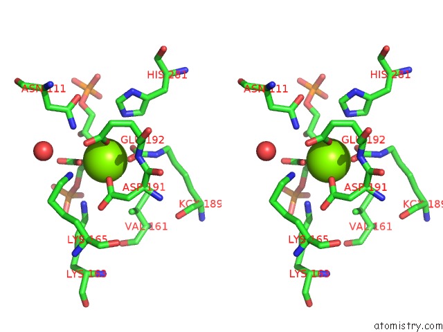
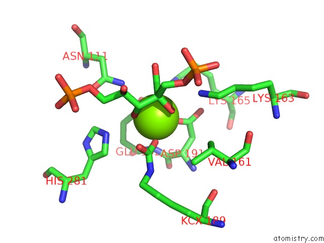
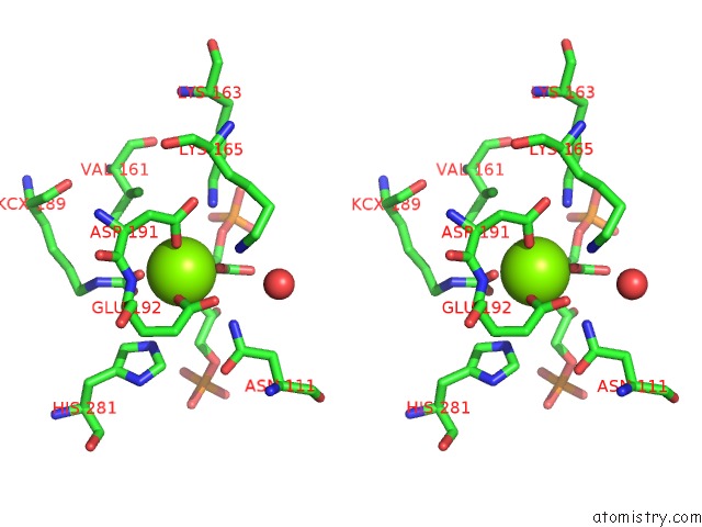
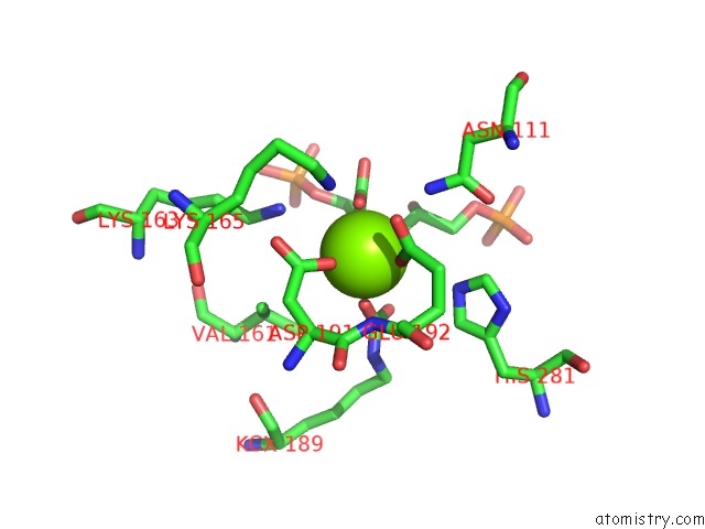
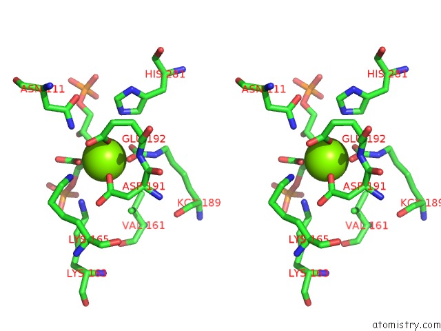
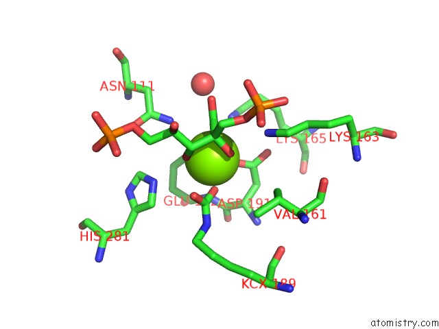
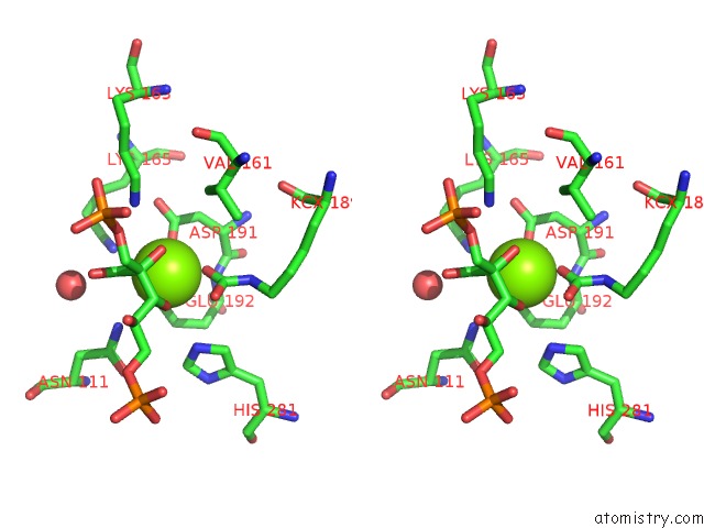
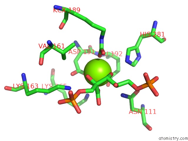
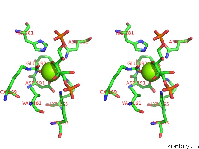
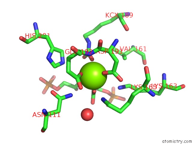
Magnesium binding site 1 out of 10 in 3kdn
Go back to
Magnesium binding site 1 out
of 10 in the Crystal Structure of Type III Rubisco SP4 Mutant Complexed with 2-Cabp
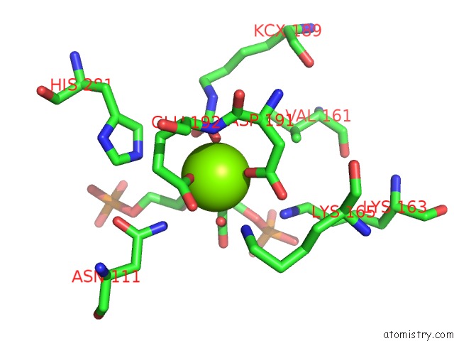
Mono view
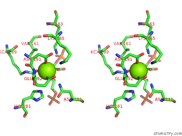
Stereo pair view
Mono view
Stereo pair view
A full contact list of Magnesium with other atoms in the Mg binding
site number 1 of Crystal Structure of Type III Rubisco SP4 Mutant Complexed with 2-Cabp within 5.0Å range:
|
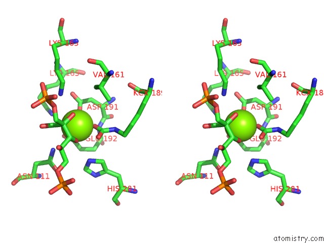
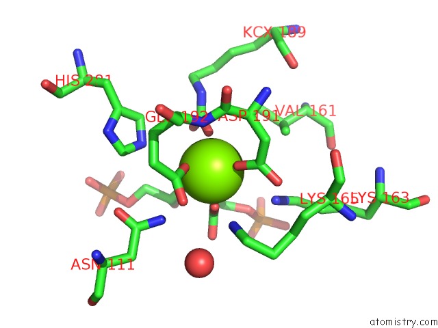
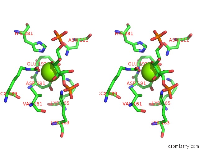
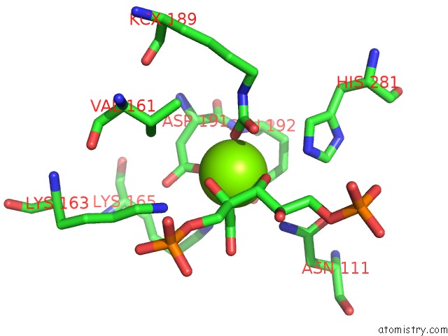
Magnesium binding site 2 out of 10 in 3kdn
Go back to
Magnesium binding site 2 out
of 10 in the Crystal Structure of Type III Rubisco SP4 Mutant Complexed with 2-Cabp
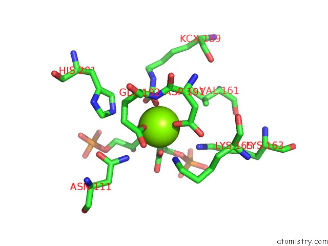
Mono view

Stereo pair view
Mono view
Stereo pair view
A full contact list of Magnesium with other atoms in the Mg binding
site number 2 of Crystal Structure of Type III Rubisco SP4 Mutant Complexed with 2-Cabp within 5.0Å range:
|
Magnesium binding site 3 out of 10 in 3kdn
Go back to
Magnesium binding site 3 out
of 10 in the Crystal Structure of Type III Rubisco SP4 Mutant Complexed with 2-Cabp
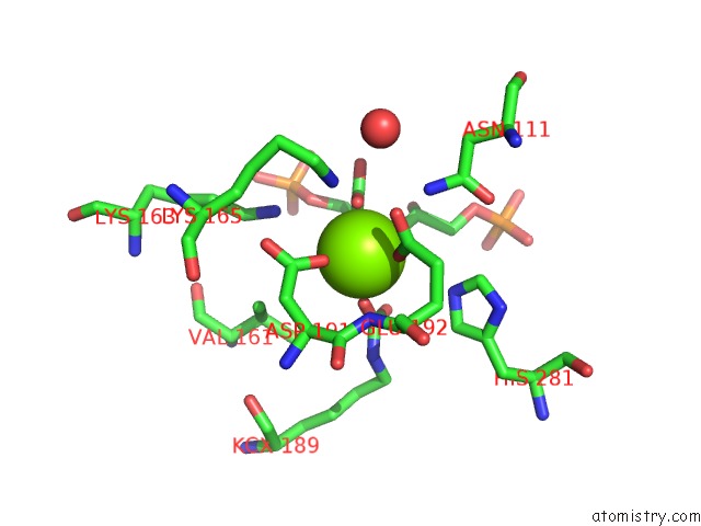
Mono view
Stereo pair view
Mono view
Stereo pair view
A full contact list of Magnesium with other atoms in the Mg binding
site number 3 of Crystal Structure of Type III Rubisco SP4 Mutant Complexed with 2-Cabp within 5.0Å range:
|
Magnesium binding site 4 out of 10 in 3kdn
Go back to
Magnesium binding site 4 out
of 10 in the Crystal Structure of Type III Rubisco SP4 Mutant Complexed with 2-Cabp
Mono view
Stereo pair view
Mono view
Stereo pair view
A full contact list of Magnesium with other atoms in the Mg binding
site number 4 of Crystal Structure of Type III Rubisco SP4 Mutant Complexed with 2-Cabp within 5.0Å range:
|
Magnesium binding site 5 out of 10 in 3kdn
Go back to
Magnesium binding site 5 out
of 10 in the Crystal Structure of Type III Rubisco SP4 Mutant Complexed with 2-Cabp
Mono view
Stereo pair view
Mono view
Stereo pair view
A full contact list of Magnesium with other atoms in the Mg binding
site number 5 of Crystal Structure of Type III Rubisco SP4 Mutant Complexed with 2-Cabp within 5.0Å range:
|
Magnesium binding site 6 out of 10 in 3kdn
Go back to
Magnesium binding site 6 out
of 10 in the Crystal Structure of Type III Rubisco SP4 Mutant Complexed with 2-Cabp
Mono view
Stereo pair view
Mono view
Stereo pair view
A full contact list of Magnesium with other atoms in the Mg binding
site number 6 of Crystal Structure of Type III Rubisco SP4 Mutant Complexed with 2-Cabp within 5.0Å range:
|
Magnesium binding site 7 out of 10 in 3kdn
Go back to
Magnesium binding site 7 out
of 10 in the Crystal Structure of Type III Rubisco SP4 Mutant Complexed with 2-Cabp
Mono view
Stereo pair view
Mono view
Stereo pair view
A full contact list of Magnesium with other atoms in the Mg binding
site number 7 of Crystal Structure of Type III Rubisco SP4 Mutant Complexed with 2-Cabp within 5.0Å range:
|
Magnesium binding site 8 out of 10 in 3kdn
Go back to
Magnesium binding site 8 out
of 10 in the Crystal Structure of Type III Rubisco SP4 Mutant Complexed with 2-Cabp
Mono view
Stereo pair view
Mono view
Stereo pair view
A full contact list of Magnesium with other atoms in the Mg binding
site number 8 of Crystal Structure of Type III Rubisco SP4 Mutant Complexed with 2-Cabp within 5.0Å range:
|
Magnesium binding site 9 out of 10 in 3kdn
Go back to
Magnesium binding site 9 out
of 10 in the Crystal Structure of Type III Rubisco SP4 Mutant Complexed with 2-Cabp
Mono view
Stereo pair view
Mono view
Stereo pair view
A full contact list of Magnesium with other atoms in the Mg binding
site number 9 of Crystal Structure of Type III Rubisco SP4 Mutant Complexed with 2-Cabp within 5.0Å range:
|
Magnesium binding site 10 out of 10 in 3kdn
Go back to
Magnesium binding site 10 out
of 10 in the Crystal Structure of Type III Rubisco SP4 Mutant Complexed with 2-Cabp
Mono view

Stereo pair view
Mono view
Stereo pair view
A full contact list of Magnesium with other atoms in the Mg binding
site number 10 of Crystal Structure of Type III Rubisco SP4 Mutant Complexed with 2-Cabp within 5.0Å range:
|
Reference:
Y.Nishitani,
S.Yoshida,
M.Fujihashi,
K.Kitagawa,
T.Doi,
H.Atomi,
T.Imanaka,
K.Miki.
Structure-Based Catalytic Optimization of A Type III Rubisco From A Hyperthermophile J.Biol.Chem. V. 285 39339 2010.
ISSN: ISSN 0021-9258
PubMed: 20926376
DOI: 10.1074/JBC.M110.147587
Page generated: Wed Aug 14 18:02:45 2024
ISSN: ISSN 0021-9258
PubMed: 20926376
DOI: 10.1074/JBC.M110.147587
Last articles
Zn in 9MJ5Zn in 9HNW
Zn in 9G0L
Zn in 9FNE
Zn in 9DZN
Zn in 9E0I
Zn in 9D32
Zn in 9DAK
Zn in 8ZXC
Zn in 8ZUF