Magnesium »
PDB 3k9l-3kk1 »
3kfu »
Magnesium in PDB 3kfu: Crystal Structure of the Transamidosome
Protein crystallography data
The structure of Crystal Structure of the Transamidosome, PDB code: 3kfu
was solved by
M.Blaise,
M.Bailly,
M.Frechin,
S.Thirup,
H.D.Becker,
D.Kern,
with X-Ray Crystallography technique. A brief refinement statistics is given in the table below:
| Resolution Low / High (Å) | 39.19 / 3.00 |
| Space group | P 1 21 1 |
| Cell size a, b, c (Å), α, β, γ (°) | 115.920, 214.000, 127.840, 90.00, 93.36, 90.00 |
| R / Rfree (%) | 19.9 / 25.2 |
Other elements in 3kfu:
The structure of Crystal Structure of the Transamidosome also contains other interesting chemical elements:
| Zinc | (Zn) | 2 atoms |
Magnesium Binding Sites:
The binding sites of Magnesium atom in the Crystal Structure of the Transamidosome
(pdb code 3kfu). This binding sites where shown within
5.0 Angstroms radius around Magnesium atom.
In total 2 binding sites of Magnesium where determined in the Crystal Structure of the Transamidosome, PDB code: 3kfu:
Jump to Magnesium binding site number: 1; 2;
In total 2 binding sites of Magnesium where determined in the Crystal Structure of the Transamidosome, PDB code: 3kfu:
Jump to Magnesium binding site number: 1; 2;
Magnesium binding site 1 out of 2 in 3kfu
Go back to
Magnesium binding site 1 out
of 2 in the Crystal Structure of the Transamidosome
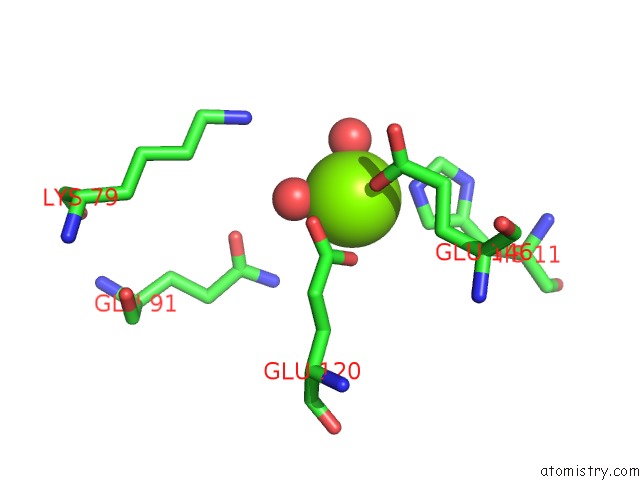
Mono view
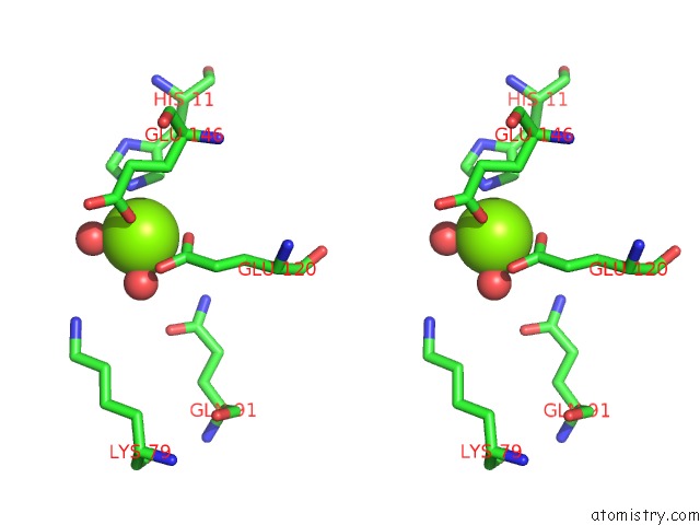
Stereo pair view
Mono view
Stereo pair view
A full contact list of Magnesium with other atoms in the Mg binding
site number 1 of Crystal Structure of the Transamidosome within 5.0Å range:
|
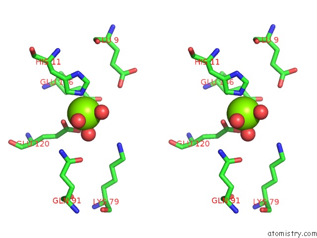
Magnesium binding site 2 out of 2 in 3kfu
Go back to
Magnesium binding site 2 out
of 2 in the Crystal Structure of the Transamidosome
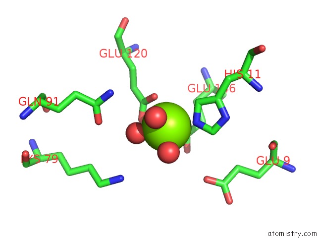
Mono view
Stereo pair view
Mono view
Stereo pair view
A full contact list of Magnesium with other atoms in the Mg binding
site number 2 of Crystal Structure of the Transamidosome within 5.0Å range:
|
Reference:
M.Blaise,
M.Bailly,
M.Frechin,
M.A.Behrens,
F.Fischer,
C.L.Oliveira,
H.D.Becker,
J.S.Pedersen,
S.Thirup,
D.Kern.
Crystal Structure of A Transfer-Ribonucleoprotein Particle That Promotes Asparagine Formation. Embo J. V. 29 3118 2010.
ISSN: ISSN 0261-4189
PubMed: 20717102
DOI: 10.1038/EMBOJ.2010.192
Page generated: Wed Aug 14 18:07:24 2024
ISSN: ISSN 0261-4189
PubMed: 20717102
DOI: 10.1038/EMBOJ.2010.192
Last articles
F in 7L07F in 7L01
F in 7L0E
F in 7L03
F in 7KYY
F in 7KYK
F in 7KZY
F in 7KZ4
F in 7KYV
F in 7KYT