Magnesium »
PDB 3k9l-3kk1 »
3kga »
Magnesium in PDB 3kga: Crystal Structure of Mapkap Kinase 2 (MK2) Complexed with A Potent 3-Aminopyrazole Atp Site Inhibitor
Enzymatic activity of Crystal Structure of Mapkap Kinase 2 (MK2) Complexed with A Potent 3-Aminopyrazole Atp Site Inhibitor
All present enzymatic activity of Crystal Structure of Mapkap Kinase 2 (MK2) Complexed with A Potent 3-Aminopyrazole Atp Site Inhibitor:
2.7.11.1;
2.7.11.1;
Protein crystallography data
The structure of Crystal Structure of Mapkap Kinase 2 (MK2) Complexed with A Potent 3-Aminopyrazole Atp Site Inhibitor, PDB code: 3kga
was solved by
M.Kroemer,
J.Velcicky,
A.Izaac,
C.Be,
C.Huppertz,
D.Pflieger,
A.Schlapbach,
C.Scheufler,
with X-Ray Crystallography technique. A brief refinement statistics is given in the table below:
| Resolution Low / High (Å) | 29.30 / 2.55 |
| Space group | P 63 2 2 |
| Cell size a, b, c (Å), α, β, γ (°) | 103.120, 103.120, 165.415, 90.00, 90.00, 120.00 |
| R / Rfree (%) | 18 / 23.5 |
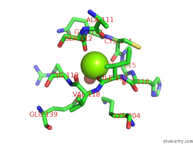
Magnesium Binding Sites:
The binding sites of Magnesium atom in the Crystal Structure of Mapkap Kinase 2 (MK2) Complexed with A Potent 3-Aminopyrazole Atp Site Inhibitor
(pdb code 3kga). This binding sites where shown within
5.0 Angstroms radius around Magnesium atom.
In total only one binding site of Magnesium was determined in the Crystal Structure of Mapkap Kinase 2 (MK2) Complexed with A Potent 3-Aminopyrazole Atp Site Inhibitor, PDB code: 3kga:
In total only one binding site of Magnesium was determined in the Crystal Structure of Mapkap Kinase 2 (MK2) Complexed with A Potent 3-Aminopyrazole Atp Site Inhibitor, PDB code: 3kga:
Magnesium binding site 1 out of 1 in 3kga
Go back to
Magnesium binding site 1 out
of 1 in the Crystal Structure of Mapkap Kinase 2 (MK2) Complexed with A Potent 3-Aminopyrazole Atp Site Inhibitor
Mono view
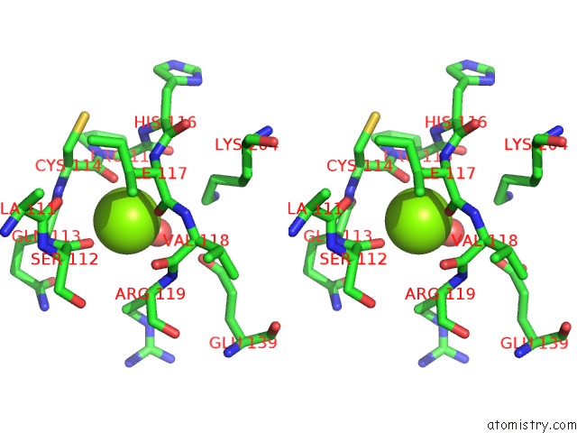
Stereo pair view
Mono view
Stereo pair view
A full contact list of Magnesium with other atoms in the Mg binding
site number 1 of Crystal Structure of Mapkap Kinase 2 (MK2) Complexed with A Potent 3-Aminopyrazole Atp Site Inhibitor within 5.0Å range:
|
Reference:
J.Velcicky,
R.Feifel,
S.Hawtin,
R.Heng,
C.Huppertz,
G.Koch,
M.Kroemer,
H.Moebitz,
L.Revesz,
C.Scheufler,
A.Schlapbach.
Novel 3-Aminopyrazole Inhibitors of Mk-2 Discovered By Scaffold Hopping Strategy. Bioorg.Med.Chem.Lett. V. 20 1293 2010.
ISSN: ISSN 0960-894X
PubMed: 20060294
DOI: 10.1016/J.BMCL.2009.10.138
Page generated: Wed Aug 14 18:07:27 2024
ISSN: ISSN 0960-894X
PubMed: 20060294
DOI: 10.1016/J.BMCL.2009.10.138
Last articles
Zn in 9MJ5Zn in 9HNW
Zn in 9G0L
Zn in 9FNE
Zn in 9DZN
Zn in 9E0I
Zn in 9D32
Zn in 9DAK
Zn in 8ZXC
Zn in 8ZUF