Magnesium »
PDB 3k9l-3kk1 »
3khq »
Magnesium in PDB 3khq: Crystal Structure of Murine Ig-Beta (CD79B) in the Monomeric Form
Protein crystallography data
The structure of Crystal Structure of Murine Ig-Beta (CD79B) in the Monomeric Form, PDB code: 3khq
was solved by
S.Radaev,
P.D.Sun,
with X-Ray Crystallography technique. A brief refinement statistics is given in the table below:
| Resolution Low / High (Å) | 39.87 / 1.70 |
| Space group | P 21 21 2 |
| Cell size a, b, c (Å), α, β, γ (°) | 35.630, 79.720, 34.320, 90.00, 90.00, 90.00 |
| R / Rfree (%) | 18.7 / 19.7 |
Magnesium Binding Sites:
The binding sites of Magnesium atom in the Crystal Structure of Murine Ig-Beta (CD79B) in the Monomeric Form
(pdb code 3khq). This binding sites where shown within
5.0 Angstroms radius around Magnesium atom.
In total only one binding site of Magnesium was determined in the Crystal Structure of Murine Ig-Beta (CD79B) in the Monomeric Form, PDB code: 3khq:
In total only one binding site of Magnesium was determined in the Crystal Structure of Murine Ig-Beta (CD79B) in the Monomeric Form, PDB code: 3khq:
Magnesium binding site 1 out of 1 in 3khq
Go back to
Magnesium binding site 1 out
of 1 in the Crystal Structure of Murine Ig-Beta (CD79B) in the Monomeric Form
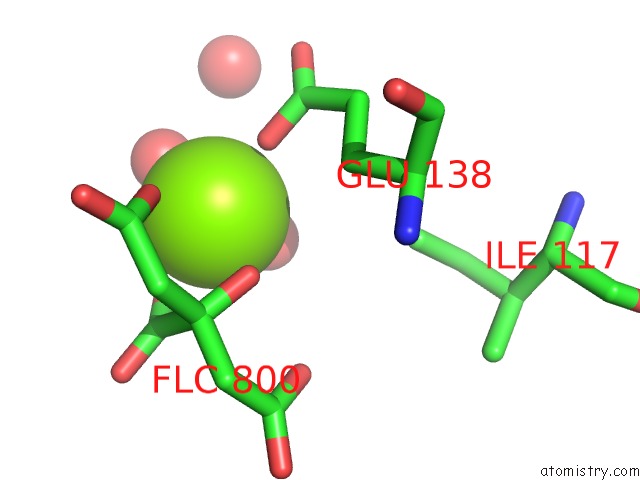
Mono view
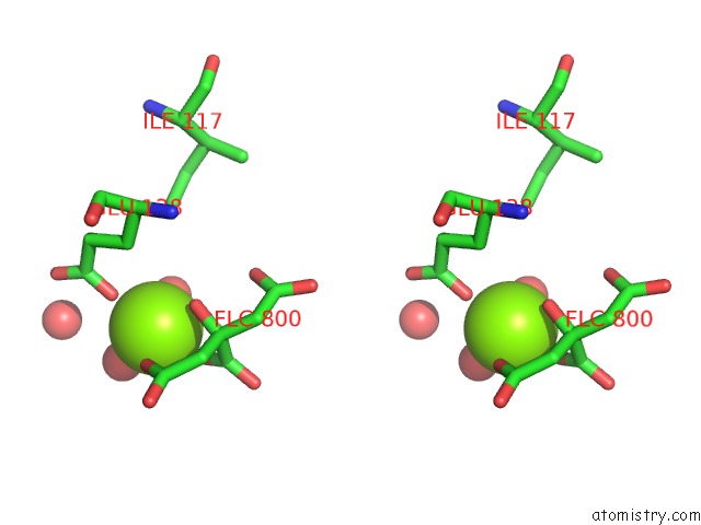
Stereo pair view
Mono view
Stereo pair view
A full contact list of Magnesium with other atoms in the Mg binding
site number 1 of Crystal Structure of Murine Ig-Beta (CD79B) in the Monomeric Form within 5.0Å range:
|
Reference:
S.Radaev,
Z.Zou,
P.Tolar,
K.Nguyen,
A.Nguyen,
P.D.Krueger,
N.Stutzman,
S.Pierce,
P.D.Sun.
Structural and Functional Studies of Igalphabeta and Its Assembly with the B Cell Antigen Receptor. Structure V. 18 934 2010.
ISSN: ISSN 0969-2126
PubMed: 20696394
DOI: 10.1016/J.STR.2010.04.019
Page generated: Wed Aug 14 18:08:10 2024
ISSN: ISSN 0969-2126
PubMed: 20696394
DOI: 10.1016/J.STR.2010.04.019
Last articles
Zn in 9J0NZn in 9J0O
Zn in 9J0P
Zn in 9FJX
Zn in 9EKB
Zn in 9C0F
Zn in 9CAH
Zn in 9CH0
Zn in 9CH3
Zn in 9CH1