Magnesium »
PDB 3k9l-3kk1 »
3kia »
Magnesium in PDB 3kia: Crystal Structure of Mannosyl-3-Phosphoglycerate Synthase From Rubrobacter Xylanophilus
Enzymatic activity of Crystal Structure of Mannosyl-3-Phosphoglycerate Synthase From Rubrobacter Xylanophilus
All present enzymatic activity of Crystal Structure of Mannosyl-3-Phosphoglycerate Synthase From Rubrobacter Xylanophilus:
2.4.1.217;
2.4.1.217;
Protein crystallography data
The structure of Crystal Structure of Mannosyl-3-Phosphoglycerate Synthase From Rubrobacter Xylanophilus, PDB code: 3kia
was solved by
S.Macedo-Ribeiro,
P.J.B.Pereira,
N.Empadinhas,
M.S.Da Costa,
with X-Ray Crystallography technique. A brief refinement statistics is given in the table below:
| Resolution Low / High (Å) | 53.70 / 2.80 |
| Space group | P 65 2 2 |
| Cell size a, b, c (Å), α, β, γ (°) | 109.010, 109.010, 313.418, 90.00, 90.00, 120.00 |
| R / Rfree (%) | 19 / 23.9 |
Other elements in 3kia:
The structure of Crystal Structure of Mannosyl-3-Phosphoglycerate Synthase From Rubrobacter Xylanophilus also contains other interesting chemical elements:
| Chlorine | (Cl) | 3 atoms |
Magnesium Binding Sites:
The binding sites of Magnesium atom in the Crystal Structure of Mannosyl-3-Phosphoglycerate Synthase From Rubrobacter Xylanophilus
(pdb code 3kia). This binding sites where shown within
5.0 Angstroms radius around Magnesium atom.
In total 3 binding sites of Magnesium where determined in the Crystal Structure of Mannosyl-3-Phosphoglycerate Synthase From Rubrobacter Xylanophilus, PDB code: 3kia:
Jump to Magnesium binding site number: 1; 2; 3;
In total 3 binding sites of Magnesium where determined in the Crystal Structure of Mannosyl-3-Phosphoglycerate Synthase From Rubrobacter Xylanophilus, PDB code: 3kia:
Jump to Magnesium binding site number: 1; 2; 3;
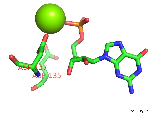
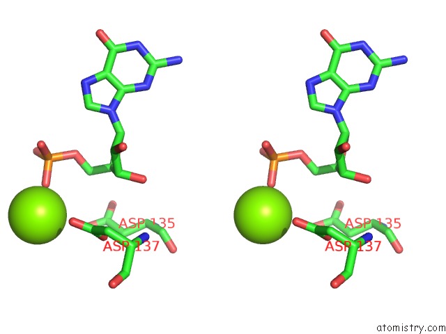
Magnesium binding site 1 out of 3 in 3kia
Go back to
Magnesium binding site 1 out
of 3 in the Crystal Structure of Mannosyl-3-Phosphoglycerate Synthase From Rubrobacter Xylanophilus
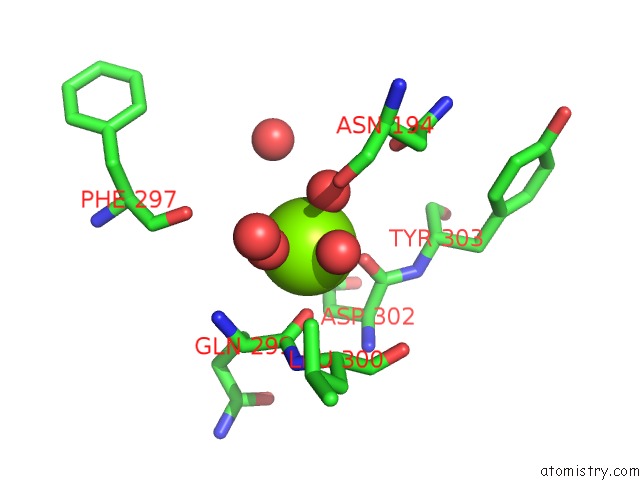
Mono view
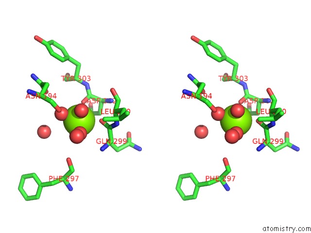
Stereo pair view
Mono view
Stereo pair view
A full contact list of Magnesium with other atoms in the Mg binding
site number 1 of Crystal Structure of Mannosyl-3-Phosphoglycerate Synthase From Rubrobacter Xylanophilus within 5.0Å range:
|
Magnesium binding site 2 out of 3 in 3kia
Go back to
Magnesium binding site 2 out
of 3 in the Crystal Structure of Mannosyl-3-Phosphoglycerate Synthase From Rubrobacter Xylanophilus
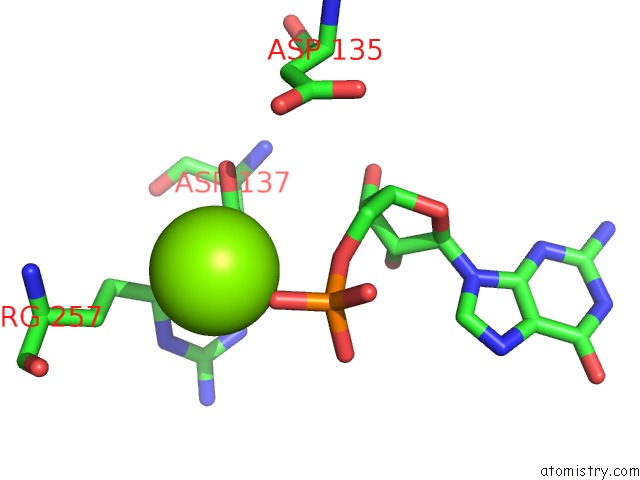
Mono view
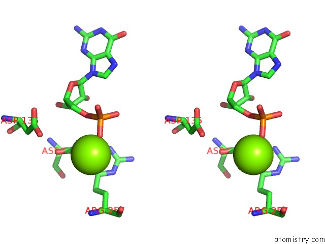
Stereo pair view
Mono view
Stereo pair view
A full contact list of Magnesium with other atoms in the Mg binding
site number 2 of Crystal Structure of Mannosyl-3-Phosphoglycerate Synthase From Rubrobacter Xylanophilus within 5.0Å range:
|
Magnesium binding site 3 out of 3 in 3kia
Go back to
Magnesium binding site 3 out
of 3 in the Crystal Structure of Mannosyl-3-Phosphoglycerate Synthase From Rubrobacter Xylanophilus
Mono view
Stereo pair view
Mono view
Stereo pair view
A full contact list of Magnesium with other atoms in the Mg binding
site number 3 of Crystal Structure of Mannosyl-3-Phosphoglycerate Synthase From Rubrobacter Xylanophilus within 5.0Å range:
|
Reference:
N.Empadinhas,
P.J.B.Pereira,
L.Albuquerque,
J.Costa,
B.Sa-Moura,
A.T.Marques,
S.Macedo-Ribeiro,
M.S.Da Costa.
Functional and Structural Characterization of A Novel Mannosyl-3-Phosphoglycerate Synthase From Rubrobacter Xylanophilus Reveals Its Dual Substrate Specificity Mol.Microbiol. V. 79 76 2011.
ISSN: ISSN 0950-382X
PubMed: 21166895
DOI: 10.1111/J.1365-2958.2010.07432.X
Page generated: Wed Aug 14 18:08:27 2024
ISSN: ISSN 0950-382X
PubMed: 21166895
DOI: 10.1111/J.1365-2958.2010.07432.X
Last articles
Zn in 9MJ5Zn in 9HNW
Zn in 9G0L
Zn in 9FNE
Zn in 9DZN
Zn in 9E0I
Zn in 9D32
Zn in 9DAK
Zn in 8ZXC
Zn in 8ZUF