Magnesium »
PDB 3kk2-3ktq »
3kms »
Magnesium in PDB 3kms: G62S Mutant of Foot-and-Mouth Disease Virus Rna-Polymerase in Complex with A Template- Primer Rna Trigonal Structure
Enzymatic activity of G62S Mutant of Foot-and-Mouth Disease Virus Rna-Polymerase in Complex with A Template- Primer Rna Trigonal Structure
All present enzymatic activity of G62S Mutant of Foot-and-Mouth Disease Virus Rna-Polymerase in Complex with A Template- Primer Rna Trigonal Structure:
2.7.7.48;
2.7.7.48;
Protein crystallography data
The structure of G62S Mutant of Foot-and-Mouth Disease Virus Rna-Polymerase in Complex with A Template- Primer Rna Trigonal Structure, PDB code: 3kms
was solved by
C.Ferrer-Orta,
N.Verdaguer,
R.Perez-Luque,
with X-Ray Crystallography technique. A brief refinement statistics is given in the table below:
| Resolution Low / High (Å) | 29.40 / 2.20 |
| Space group | P 32 2 1 |
| Cell size a, b, c (Å), α, β, γ (°) | 93.951, 93.951, 99.997, 90.00, 90.00, 120.00 |
| R / Rfree (%) | 23.6 / 26.6 |
Magnesium Binding Sites:
The binding sites of Magnesium atom in the G62S Mutant of Foot-and-Mouth Disease Virus Rna-Polymerase in Complex with A Template- Primer Rna Trigonal Structure
(pdb code 3kms). This binding sites where shown within
5.0 Angstroms radius around Magnesium atom.
In total only one binding site of Magnesium was determined in the G62S Mutant of Foot-and-Mouth Disease Virus Rna-Polymerase in Complex with A Template- Primer Rna Trigonal Structure, PDB code: 3kms:
In total only one binding site of Magnesium was determined in the G62S Mutant of Foot-and-Mouth Disease Virus Rna-Polymerase in Complex with A Template- Primer Rna Trigonal Structure, PDB code: 3kms:
Magnesium binding site 1 out of 1 in 3kms
Go back to
Magnesium binding site 1 out
of 1 in the G62S Mutant of Foot-and-Mouth Disease Virus Rna-Polymerase in Complex with A Template- Primer Rna Trigonal Structure
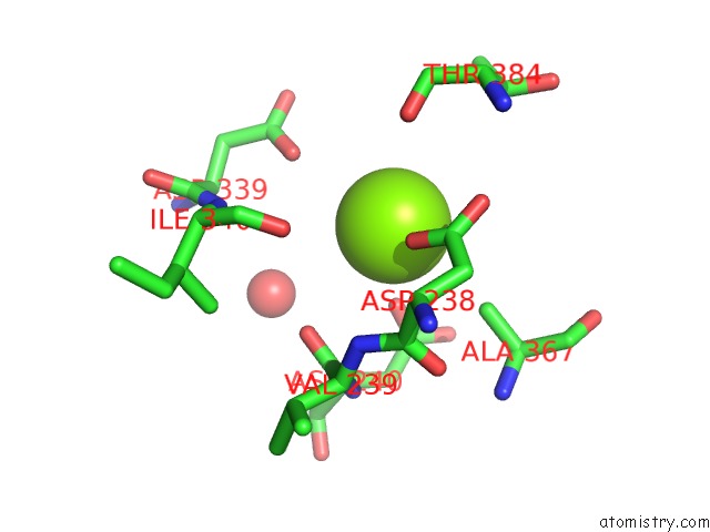
Mono view
Stereo pair view
Mono view
Stereo pair view
A full contact list of Magnesium with other atoms in the Mg binding
site number 1 of G62S Mutant of Foot-and-Mouth Disease Virus Rna-Polymerase in Complex with A Template- Primer Rna Trigonal Structure within 5.0Å range:
|
Reference:
C.Ferrer-Orta,
M.Sierra,
R.Agudo,
I.De La Higuera,
A.Arias,
R.Perez-Luque,
C.Escarmis,
E.Domingo,
N.Verdaguer.
Structure of Foot-and-Mouth Disease Virus Mutant Polymerases with Reduced Sensitivity to Ribavirin J.Virol. V. 84 6188 2010.
ISSN: ISSN 0022-538X
PubMed: 20392853
DOI: 10.1128/JVI.02420-09
Page generated: Wed Aug 14 18:11:17 2024
ISSN: ISSN 0022-538X
PubMed: 20392853
DOI: 10.1128/JVI.02420-09
Last articles
Zn in 9J0NZn in 9J0O
Zn in 9J0P
Zn in 9FJX
Zn in 9EKB
Zn in 9C0F
Zn in 9CAH
Zn in 9CH0
Zn in 9CH3
Zn in 9CH1