Magnesium »
PDB 3kk2-3ktq »
3ksa »
Magnesium in PDB 3ksa: Detailed Structural Insight Into the Dna Cleavage Complex of Type Iia Topoisomerases (Cleaved Form)
Protein crystallography data
The structure of Detailed Structural Insight Into the Dna Cleavage Complex of Type Iia Topoisomerases (Cleaved Form), PDB code: 3ksa
was solved by
I.Laponogov,
X.-S.Pan,
D.A.Veselkov,
K.E.Mcauley,
L.M.Fisher,
M.R.Sanderson,
with X-Ray Crystallography technique. A brief refinement statistics is given in the table below:
| Resolution Low / High (Å) | 29.40 / 3.30 |
| Space group | P 32 |
| Cell size a, b, c (Å), α, β, γ (°) | 116.911, 116.911, 183.759, 90.00, 90.00, 120.00 |
| R / Rfree (%) | 17.6 / 21.8 |
Magnesium Binding Sites:
The binding sites of Magnesium atom in the Detailed Structural Insight Into the Dna Cleavage Complex of Type Iia Topoisomerases (Cleaved Form)
(pdb code 3ksa). This binding sites where shown within
5.0 Angstroms radius around Magnesium atom.
In total 2 binding sites of Magnesium where determined in the Detailed Structural Insight Into the Dna Cleavage Complex of Type Iia Topoisomerases (Cleaved Form), PDB code: 3ksa:
Jump to Magnesium binding site number: 1; 2;
In total 2 binding sites of Magnesium where determined in the Detailed Structural Insight Into the Dna Cleavage Complex of Type Iia Topoisomerases (Cleaved Form), PDB code: 3ksa:
Jump to Magnesium binding site number: 1; 2;
Magnesium binding site 1 out of 2 in 3ksa
Go back to
Magnesium binding site 1 out
of 2 in the Detailed Structural Insight Into the Dna Cleavage Complex of Type Iia Topoisomerases (Cleaved Form)
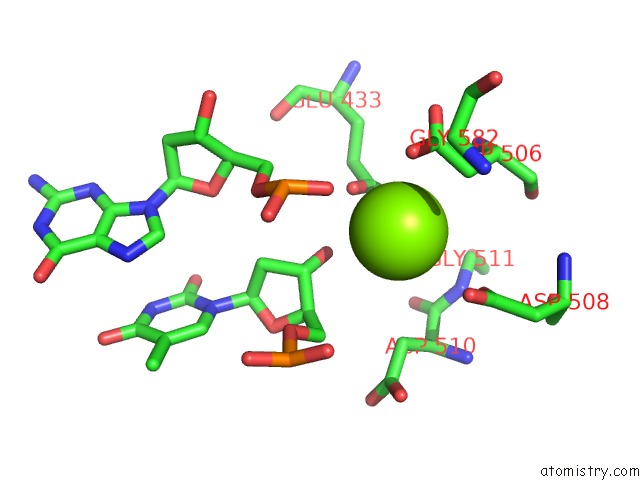
Mono view
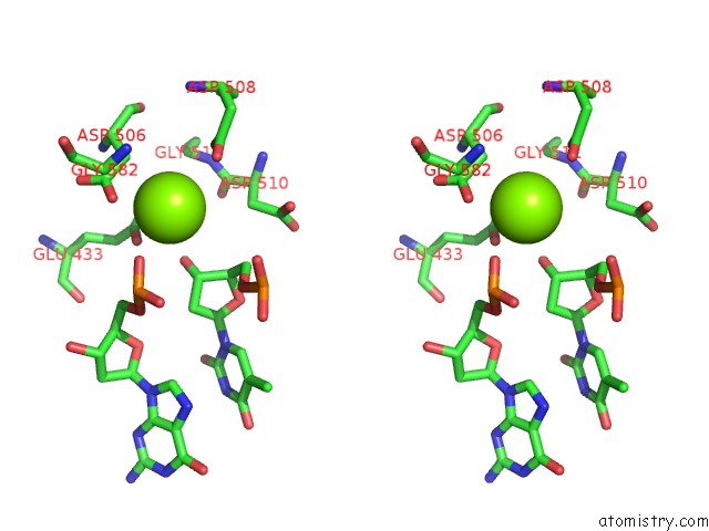
Stereo pair view
Mono view
Stereo pair view
A full contact list of Magnesium with other atoms in the Mg binding
site number 1 of Detailed Structural Insight Into the Dna Cleavage Complex of Type Iia Topoisomerases (Cleaved Form) within 5.0Å range:
|
Magnesium binding site 2 out of 2 in 3ksa
Go back to
Magnesium binding site 2 out
of 2 in the Detailed Structural Insight Into the Dna Cleavage Complex of Type Iia Topoisomerases (Cleaved Form)
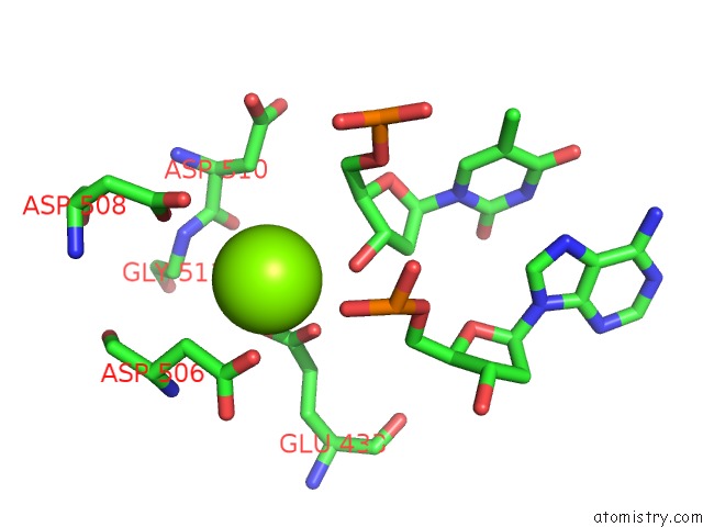
Mono view
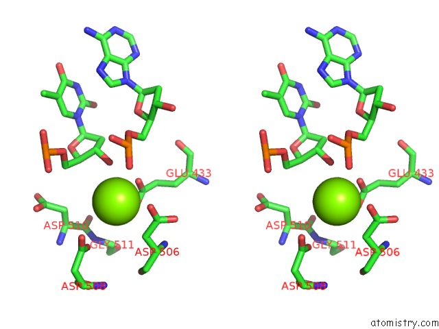
Stereo pair view
Mono view
Stereo pair view
A full contact list of Magnesium with other atoms in the Mg binding
site number 2 of Detailed Structural Insight Into the Dna Cleavage Complex of Type Iia Topoisomerases (Cleaved Form) within 5.0Å range:
|
Reference:
I.Laponogov,
X.-S.Pan,
D.A.Veselkov,
K.E.Mcauley,
L.M.Fisher,
M.R.Sanderson.
Structural Basis of Gate-Dna Breakage and Resealing By Type II Topoisomerases Plos One V. 5 E1133 2010.
ISSN: ESSN 1932-6203
PubMed: 20596531
DOI: 10.1371/JOURNAL.PONE.0011338
Page generated: Wed Aug 14 18:15:01 2024
ISSN: ESSN 1932-6203
PubMed: 20596531
DOI: 10.1371/JOURNAL.PONE.0011338
Last articles
Ca in 5TNCCa in 5TP9
Ca in 5TP0
Ca in 5TMN
Ca in 5TLI
Ca in 5TLN
Ca in 5TH6
Ca in 5TL8
Ca in 5TIW
Ca in 5TJ6