Magnesium »
PDB 3l9b-3lop »
3las »
Magnesium in PDB 3las: Crystal Structure of Carbonic Anhydrase From Streptococcus Mutans to 1.4 Angstrom Resolution
Enzymatic activity of Crystal Structure of Carbonic Anhydrase From Streptococcus Mutans to 1.4 Angstrom Resolution
All present enzymatic activity of Crystal Structure of Carbonic Anhydrase From Streptococcus Mutans to 1.4 Angstrom Resolution:
4.2.1.1;
4.2.1.1;
Protein crystallography data
The structure of Crystal Structure of Carbonic Anhydrase From Streptococcus Mutans to 1.4 Angstrom Resolution, PDB code: 3las
was solved by
L.-L.Ma,
K.-T.Wang,
X.Liu,
X.-D.Su,
with X-Ray Crystallography technique. A brief refinement statistics is given in the table below:
| Resolution Low / High (Å) | 20.00 / 1.40 |
| Space group | H 3 |
| Cell size a, b, c (Å), α, β, γ (°) | 80.949, 80.949, 128.949, 90.00, 90.00, 120.00 |
| R / Rfree (%) | 15.4 / 18 |
Other elements in 3las:
The structure of Crystal Structure of Carbonic Anhydrase From Streptococcus Mutans to 1.4 Angstrom Resolution also contains other interesting chemical elements:
| Zinc | (Zn) | 2 atoms |
Magnesium Binding Sites:
The binding sites of Magnesium atom in the Crystal Structure of Carbonic Anhydrase From Streptococcus Mutans to 1.4 Angstrom Resolution
(pdb code 3las). This binding sites where shown within
5.0 Angstroms radius around Magnesium atom.
In total 2 binding sites of Magnesium where determined in the Crystal Structure of Carbonic Anhydrase From Streptococcus Mutans to 1.4 Angstrom Resolution, PDB code: 3las:
Jump to Magnesium binding site number: 1; 2;
In total 2 binding sites of Magnesium where determined in the Crystal Structure of Carbonic Anhydrase From Streptococcus Mutans to 1.4 Angstrom Resolution, PDB code: 3las:
Jump to Magnesium binding site number: 1; 2;
Magnesium binding site 1 out of 2 in 3las
Go back to
Magnesium binding site 1 out
of 2 in the Crystal Structure of Carbonic Anhydrase From Streptococcus Mutans to 1.4 Angstrom Resolution
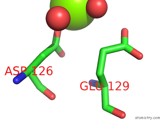
Mono view
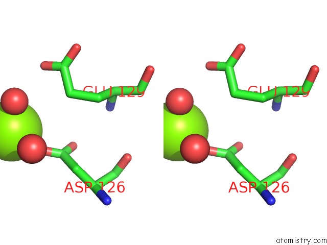
Stereo pair view
Mono view
Stereo pair view
A full contact list of Magnesium with other atoms in the Mg binding
site number 1 of Crystal Structure of Carbonic Anhydrase From Streptococcus Mutans to 1.4 Angstrom Resolution within 5.0Å range:
|
Magnesium binding site 2 out of 2 in 3las
Go back to
Magnesium binding site 2 out
of 2 in the Crystal Structure of Carbonic Anhydrase From Streptococcus Mutans to 1.4 Angstrom Resolution
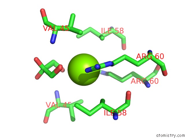
Mono view
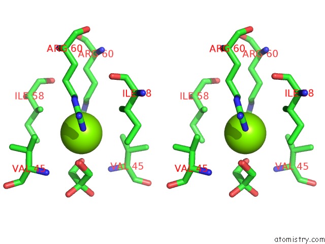
Stereo pair view
Mono view
Stereo pair view
A full contact list of Magnesium with other atoms in the Mg binding
site number 2 of Crystal Structure of Carbonic Anhydrase From Streptococcus Mutans to 1.4 Angstrom Resolution within 5.0Å range:
|
Reference:
L.-L.Ma,
K.-T.Wang,
X.Liu,
X.-D.Su.
Crystal Structure of Carbonic Anhydrase From Streptococcus Mutans to 1.4 Angstrom Resolution To Be Published.
Page generated: Wed Aug 14 18:29:08 2024
Last articles
Ca in 5UBOCa in 5U95
Ca in 5UBA
Ca in 5UB5
Ca in 5U8O
Ca in 5U6J
Ca in 5U5N
Ca in 5U5I
Ca in 5U3K
Ca in 5U3E