Magnesium »
PDB 3lpl-3m1i »
3lqq »
Magnesium in PDB 3lqq: Structure of the Ced-4 Apoptosome
Protein crystallography data
The structure of Structure of the Ced-4 Apoptosome, PDB code: 3lqq
was solved by
S.Qi,
Y.Pang,
Y.Shi,
N.Yan,
Q.Liu,
with X-Ray Crystallography technique. A brief refinement statistics is given in the table below:
| Resolution Low / High (Å) | 28.66 / 3.53 |
| Space group | I 4 2 2 |
| Cell size a, b, c (Å), α, β, γ (°) | 181.640, 181.640, 203.190, 90.00, 90.00, 90.00 |
| R / Rfree (%) | 27.5 / 31.1 |
Magnesium Binding Sites:
The binding sites of Magnesium atom in the Structure of the Ced-4 Apoptosome
(pdb code 3lqq). This binding sites where shown within
5.0 Angstroms radius around Magnesium atom.
In total 2 binding sites of Magnesium where determined in the Structure of the Ced-4 Apoptosome, PDB code: 3lqq:
Jump to Magnesium binding site number: 1; 2;
In total 2 binding sites of Magnesium where determined in the Structure of the Ced-4 Apoptosome, PDB code: 3lqq:
Jump to Magnesium binding site number: 1; 2;
Magnesium binding site 1 out of 2 in 3lqq
Go back to
Magnesium binding site 1 out
of 2 in the Structure of the Ced-4 Apoptosome
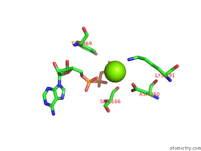
Mono view

Stereo pair view
Mono view
Stereo pair view
A full contact list of Magnesium with other atoms in the Mg binding
site number 1 of Structure of the Ced-4 Apoptosome within 5.0Å range:
|
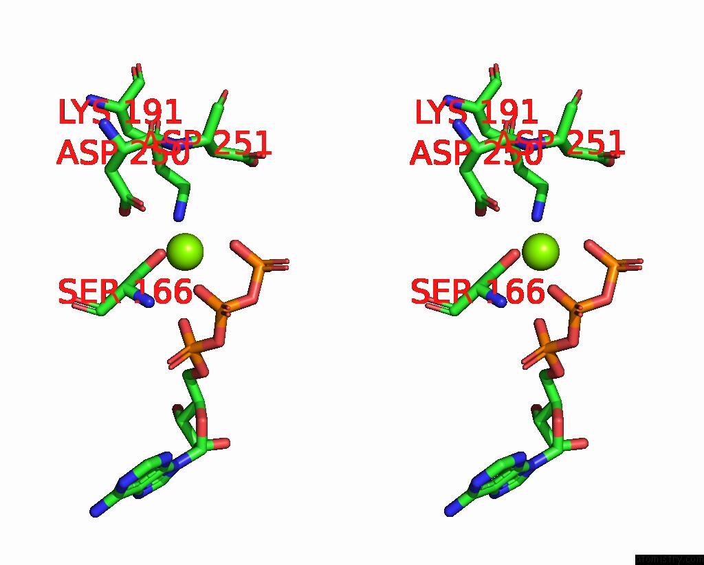
Magnesium binding site 2 out of 2 in 3lqq
Go back to
Magnesium binding site 2 out
of 2 in the Structure of the Ced-4 Apoptosome

Mono view
Stereo pair view
Mono view
Stereo pair view
A full contact list of Magnesium with other atoms in the Mg binding
site number 2 of Structure of the Ced-4 Apoptosome within 5.0Å range:
|
Reference:
S.Qi,
Y.Pang,
Q.Hu,
Q.Liu,
H.Li,
Y.Zhou,
T.He,
Q.Liang,
Y.Liu,
X.Yuan,
G.Luo,
H.Li,
J.Wang,
N.Yan,
Y.Shi.
Crystal Structure of the Caenorhabditis Elegans Apoptosome Reveals An Octameric Assembly of Ced-4. Cell(Cambridge,Mass.) V. 141 446 2010.
ISSN: ISSN 0092-8674
PubMed: 20434985
DOI: 10.1016/J.CELL.2010.03.017
Page generated: Wed Aug 14 18:37:50 2024
ISSN: ISSN 0092-8674
PubMed: 20434985
DOI: 10.1016/J.CELL.2010.03.017
Last articles
Zn in 9MJ5Zn in 9HNW
Zn in 9G0L
Zn in 9FNE
Zn in 9DZN
Zn in 9E0I
Zn in 9D32
Zn in 9DAK
Zn in 8ZXC
Zn in 8ZUF