Magnesium »
PDB 3m1v-3mfh »
3m6m »
Magnesium in PDB 3m6m: Crystal Structure of Rpff Complexed with Rec Domain of Rpfc
Enzymatic activity of Crystal Structure of Rpff Complexed with Rec Domain of Rpfc
All present enzymatic activity of Crystal Structure of Rpff Complexed with Rec Domain of Rpfc:
2.7.13.3;
2.7.13.3;
Protein crystallography data
The structure of Crystal Structure of Rpff Complexed with Rec Domain of Rpfc, PDB code: 3m6m
was solved by
Z.Cheng,
S.C.Lim,
R.Qamra,
H.Song,
with X-Ray Crystallography technique. A brief refinement statistics is given in the table below:
| Resolution Low / High (Å) | 20.00 / 2.50 |
| Space group | P 65 |
| Cell size a, b, c (Å), α, β, γ (°) | 130.913, 130.913, 156.471, 90.00, 90.00, 120.00 |
| R / Rfree (%) | 25 / 27.1 |
Other elements in 3m6m:
The structure of Crystal Structure of Rpff Complexed with Rec Domain of Rpfc also contains other interesting chemical elements:
| Iodine | (I) | 14 atoms |
Magnesium Binding Sites:
The binding sites of Magnesium atom in the Crystal Structure of Rpff Complexed with Rec Domain of Rpfc
(pdb code 3m6m). This binding sites where shown within
5.0 Angstroms radius around Magnesium atom.
In total 3 binding sites of Magnesium where determined in the Crystal Structure of Rpff Complexed with Rec Domain of Rpfc, PDB code: 3m6m:
Jump to Magnesium binding site number: 1; 2; 3;
In total 3 binding sites of Magnesium where determined in the Crystal Structure of Rpff Complexed with Rec Domain of Rpfc, PDB code: 3m6m:
Jump to Magnesium binding site number: 1; 2; 3;
Magnesium binding site 1 out of 3 in 3m6m
Go back to
Magnesium binding site 1 out
of 3 in the Crystal Structure of Rpff Complexed with Rec Domain of Rpfc
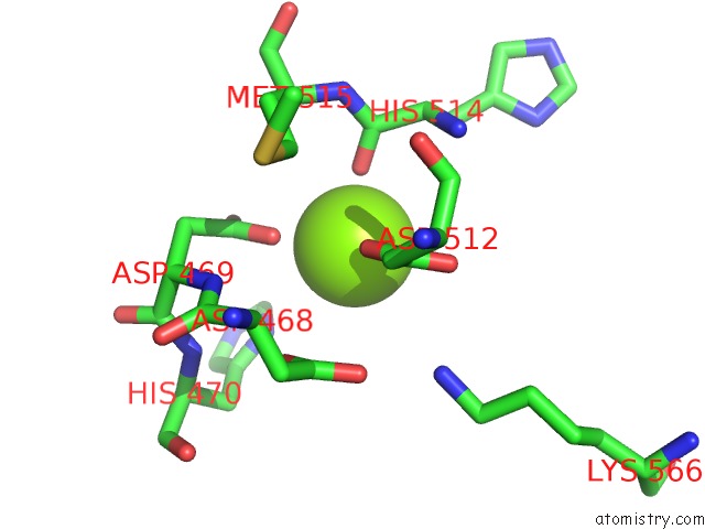
Mono view
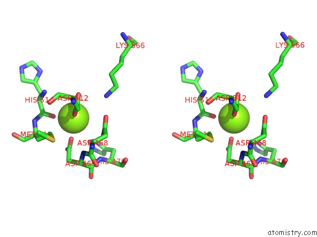
Stereo pair view
Mono view
Stereo pair view
A full contact list of Magnesium with other atoms in the Mg binding
site number 1 of Crystal Structure of Rpff Complexed with Rec Domain of Rpfc within 5.0Å range:
|
Magnesium binding site 2 out of 3 in 3m6m
Go back to
Magnesium binding site 2 out
of 3 in the Crystal Structure of Rpff Complexed with Rec Domain of Rpfc

Mono view

Stereo pair view
Mono view
Stereo pair view
A full contact list of Magnesium with other atoms in the Mg binding
site number 2 of Crystal Structure of Rpff Complexed with Rec Domain of Rpfc within 5.0Å range:
|
Magnesium binding site 3 out of 3 in 3m6m
Go back to
Magnesium binding site 3 out
of 3 in the Crystal Structure of Rpff Complexed with Rec Domain of Rpfc

Mono view

Stereo pair view
Mono view
Stereo pair view
A full contact list of Magnesium with other atoms in the Mg binding
site number 3 of Crystal Structure of Rpff Complexed with Rec Domain of Rpfc within 5.0Å range:
|
Reference:
Z.Cheng,
Y.W.He,
S.C.Lim,
R.Qamra,
M.A.Walsh,
L.H.Zhang,
H.Song.
Structural Basis of the Sensor-Synthase Interaction in Autoinduction of the Quorum Sensing Signal Dsf Biosynthesis Structure V. 18 1199 2010.
ISSN: ISSN 0969-2126
PubMed: 20826346
DOI: 10.1016/J.STR.2010.06.011
Page generated: Wed Aug 14 19:11:07 2024
ISSN: ISSN 0969-2126
PubMed: 20826346
DOI: 10.1016/J.STR.2010.06.011
Last articles
Zn in 9J0NZn in 9J0O
Zn in 9J0P
Zn in 9FJX
Zn in 9EKB
Zn in 9C0F
Zn in 9CAH
Zn in 9CH0
Zn in 9CH3
Zn in 9CH1