Magnesium »
PDB 3mfi-3mv0 »
3mjh »
Magnesium in PDB 3mjh: Crystal Structure of Human RAB5A in Complex with the C2H2 Zinc Finger of EEA1
Enzymatic activity of Crystal Structure of Human RAB5A in Complex with the C2H2 Zinc Finger of EEA1
All present enzymatic activity of Crystal Structure of Human RAB5A in Complex with the C2H2 Zinc Finger of EEA1:
3.6.5.2;
3.6.5.2;
Protein crystallography data
The structure of Crystal Structure of Human RAB5A in Complex with the C2H2 Zinc Finger of EEA1, PDB code: 3mjh
was solved by
A.K.Mishra,
S.Eathiraj,
D.G.Lambright,
with X-Ray Crystallography technique. A brief refinement statistics is given in the table below:
| Resolution Low / High (Å) | 20.00 / 2.03 |
| Space group | P 21 21 21 |
| Cell size a, b, c (Å), α, β, γ (°) | 46.412, 80.398, 103.495, 90.00, 90.00, 90.00 |
| R / Rfree (%) | 19.2 / 26 |
Other elements in 3mjh:
The structure of Crystal Structure of Human RAB5A in Complex with the C2H2 Zinc Finger of EEA1 also contains other interesting chemical elements:
| Zinc | (Zn) | 2 atoms |
Magnesium Binding Sites:
The binding sites of Magnesium atom in the Crystal Structure of Human RAB5A in Complex with the C2H2 Zinc Finger of EEA1
(pdb code 3mjh). This binding sites where shown within
5.0 Angstroms radius around Magnesium atom.
In total 2 binding sites of Magnesium where determined in the Crystal Structure of Human RAB5A in Complex with the C2H2 Zinc Finger of EEA1, PDB code: 3mjh:
Jump to Magnesium binding site number: 1; 2;
In total 2 binding sites of Magnesium where determined in the Crystal Structure of Human RAB5A in Complex with the C2H2 Zinc Finger of EEA1, PDB code: 3mjh:
Jump to Magnesium binding site number: 1; 2;
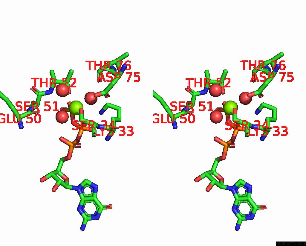
Magnesium binding site 1 out of 2 in 3mjh
Go back to
Magnesium binding site 1 out
of 2 in the Crystal Structure of Human RAB5A in Complex with the C2H2 Zinc Finger of EEA1

Mono view

Stereo pair view
Mono view
Stereo pair view
A full contact list of Magnesium with other atoms in the Mg binding
site number 1 of Crystal Structure of Human RAB5A in Complex with the C2H2 Zinc Finger of EEA1 within 5.0Å range:
|
Magnesium binding site 2 out of 2 in 3mjh
Go back to
Magnesium binding site 2 out
of 2 in the Crystal Structure of Human RAB5A in Complex with the C2H2 Zinc Finger of EEA1

Mono view
Stereo pair view
Mono view
Stereo pair view
A full contact list of Magnesium with other atoms in the Mg binding
site number 2 of Crystal Structure of Human RAB5A in Complex with the C2H2 Zinc Finger of EEA1 within 5.0Å range:
|
Reference:
A.Mishra,
S.Eathiraj,
S.Corvera,
D.G.Lambright.
Structural Basis For Rab Gtpase Recognition and Endosome Tethering By the C2H2 Zinc Finger of Early Endosomal Autoantigen 1 (EEA1). Proc.Natl.Acad.Sci.Usa V. 107 10866 2010.
ISSN: ISSN 0027-8424
PubMed: 20534488
DOI: 10.1073/PNAS.1000843107
Page generated: Wed Aug 14 19:19:45 2024
ISSN: ISSN 0027-8424
PubMed: 20534488
DOI: 10.1073/PNAS.1000843107
Last articles
Fe in 2YXOFe in 2YRS
Fe in 2YXC
Fe in 2YNM
Fe in 2YVJ
Fe in 2YP1
Fe in 2YU2
Fe in 2YU1
Fe in 2YQB
Fe in 2YOO