Magnesium »
PDB 3mfi-3mv0 »
3mk1 »
Magnesium in PDB 3mk1: Refinement of Placental Alkaline Phosphatase Complexed with Nitrophenyl
Enzymatic activity of Refinement of Placental Alkaline Phosphatase Complexed with Nitrophenyl
All present enzymatic activity of Refinement of Placental Alkaline Phosphatase Complexed with Nitrophenyl:
3.1.3.1;
3.1.3.1;
Protein crystallography data
The structure of Refinement of Placental Alkaline Phosphatase Complexed with Nitrophenyl, PDB code: 3mk1
was solved by
B.Stec,
A.Cheltsov,
J.L.Millan,
with X-Ray Crystallography technique. A brief refinement statistics is given in the table below:
| Resolution Low / High (Å) | 70.01 / 1.57 |
| Space group | C 2 2 21 |
| Cell size a, b, c (Å), α, β, γ (°) | 88.346, 114.573, 106.568, 90.00, 90.00, 90.00 |
| R / Rfree (%) | 13.9 / 17.8 |
Other elements in 3mk1:
The structure of Refinement of Placental Alkaline Phosphatase Complexed with Nitrophenyl also contains other interesting chemical elements:
| Zinc | (Zn) | 3 atoms |
| Calcium | (Ca) | 1 atom |
| Sodium | (Na) | 1 atom |
Magnesium Binding Sites:
The binding sites of Magnesium atom in the Refinement of Placental Alkaline Phosphatase Complexed with Nitrophenyl
(pdb code 3mk1). This binding sites where shown within
5.0 Angstroms radius around Magnesium atom.
In total only one binding site of Magnesium was determined in the Refinement of Placental Alkaline Phosphatase Complexed with Nitrophenyl, PDB code: 3mk1:
In total only one binding site of Magnesium was determined in the Refinement of Placental Alkaline Phosphatase Complexed with Nitrophenyl, PDB code: 3mk1:
Magnesium binding site 1 out of 1 in 3mk1
Go back to
Magnesium binding site 1 out
of 1 in the Refinement of Placental Alkaline Phosphatase Complexed with Nitrophenyl
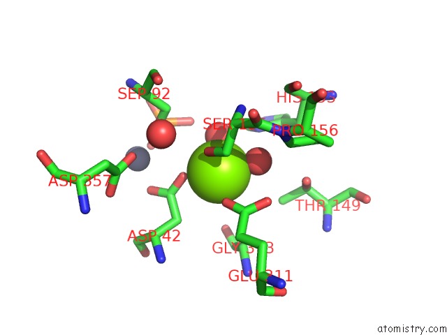
Mono view
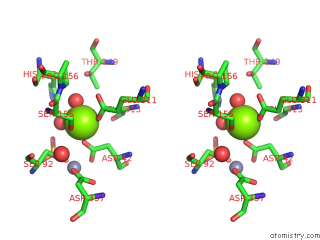
Stereo pair view
Mono view
Stereo pair view
A full contact list of Magnesium with other atoms in the Mg binding
site number 1 of Refinement of Placental Alkaline Phosphatase Complexed with Nitrophenyl within 5.0Å range:
|
Reference:
B.Stec,
A.Cheltsov,
J.L.Millan.
Refined Structures of Placental Alkaline Phosphatase Show A Consistent Pattern of Interactions at the Peripheral Site. Acta Crystallogr.,Sect.F V. 66 866 2010.
ISSN: ESSN 1744-3091
PubMed: 20693656
DOI: 10.1107/S1744309110019767
Page generated: Wed Aug 14 19:20:44 2024
ISSN: ESSN 1744-3091
PubMed: 20693656
DOI: 10.1107/S1744309110019767
Last articles
Cl in 7XO8Cl in 7XOV
Cl in 7XO7
Cl in 7XO9
Cl in 7XJO
Cl in 7XNC
Cl in 7XLP
Cl in 7XLI
Cl in 7XL9
Cl in 7XJF