Magnesium »
PDB 3mv1-3n8b »
3n6h »
Magnesium in PDB 3n6h: Crystal Structure of Mandelate Racemase/Muconate Lactonizing Protein From Actinobacillus Succinogenes 130Z Complexed with Magnesium/Sulfate
Protein crystallography data
The structure of Crystal Structure of Mandelate Racemase/Muconate Lactonizing Protein From Actinobacillus Succinogenes 130Z Complexed with Magnesium/Sulfate, PDB code: 3n6h
was solved by
V.N.Malashkevich,
Y.Patskovsky,
R.Toro,
J.M.Sauder,
S.K.Burley,
S.C.Almo,
New York Sgx Research Center For Structural Genomics (Nysgxrc),
with X-Ray Crystallography technique. A brief refinement statistics is given in the table below:
| Resolution Low / High (Å) | 20.00 / 2.30 |
| Space group | P 1 21 1 |
| Cell size a, b, c (Å), α, β, γ (°) | 97.397, 85.884, 112.424, 90.00, 96.03, 90.00 |
| R / Rfree (%) | 21.9 / 27.8 |
Other elements in 3n6h:
The structure of Crystal Structure of Mandelate Racemase/Muconate Lactonizing Protein From Actinobacillus Succinogenes 130Z Complexed with Magnesium/Sulfate also contains other interesting chemical elements:
| Chlorine | (Cl) | 1 atom |
Magnesium Binding Sites:
The binding sites of Magnesium atom in the Crystal Structure of Mandelate Racemase/Muconate Lactonizing Protein From Actinobacillus Succinogenes 130Z Complexed with Magnesium/Sulfate
(pdb code 3n6h). This binding sites where shown within
5.0 Angstroms radius around Magnesium atom.
In total 4 binding sites of Magnesium where determined in the Crystal Structure of Mandelate Racemase/Muconate Lactonizing Protein From Actinobacillus Succinogenes 130Z Complexed with Magnesium/Sulfate, PDB code: 3n6h:
Jump to Magnesium binding site number: 1; 2; 3; 4;
In total 4 binding sites of Magnesium where determined in the Crystal Structure of Mandelate Racemase/Muconate Lactonizing Protein From Actinobacillus Succinogenes 130Z Complexed with Magnesium/Sulfate, PDB code: 3n6h:
Jump to Magnesium binding site number: 1; 2; 3; 4;
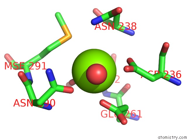
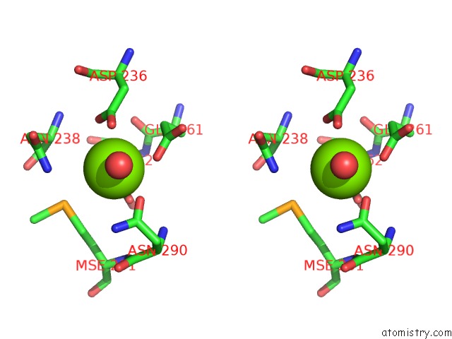
Magnesium binding site 1 out of 4 in 3n6h
Go back to
Magnesium binding site 1 out
of 4 in the Crystal Structure of Mandelate Racemase/Muconate Lactonizing Protein From Actinobacillus Succinogenes 130Z Complexed with Magnesium/Sulfate
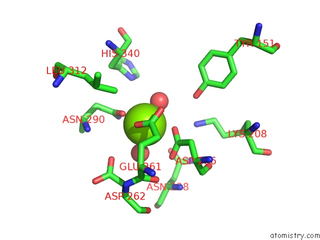
Mono view
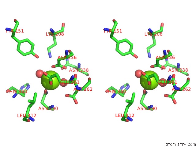
Stereo pair view
Mono view
Stereo pair view
A full contact list of Magnesium with other atoms in the Mg binding
site number 1 of Crystal Structure of Mandelate Racemase/Muconate Lactonizing Protein From Actinobacillus Succinogenes 130Z Complexed with Magnesium/Sulfate within 5.0Å range:
|
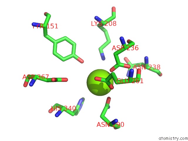
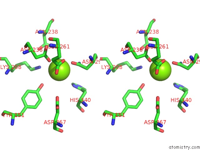
Magnesium binding site 2 out of 4 in 3n6h
Go back to
Magnesium binding site 2 out
of 4 in the Crystal Structure of Mandelate Racemase/Muconate Lactonizing Protein From Actinobacillus Succinogenes 130Z Complexed with Magnesium/Sulfate
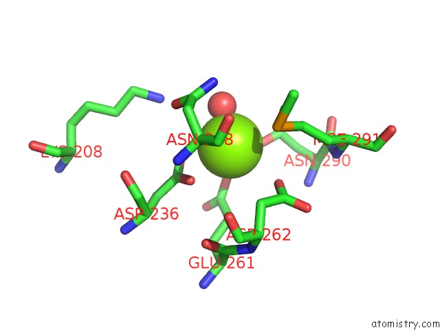
Mono view
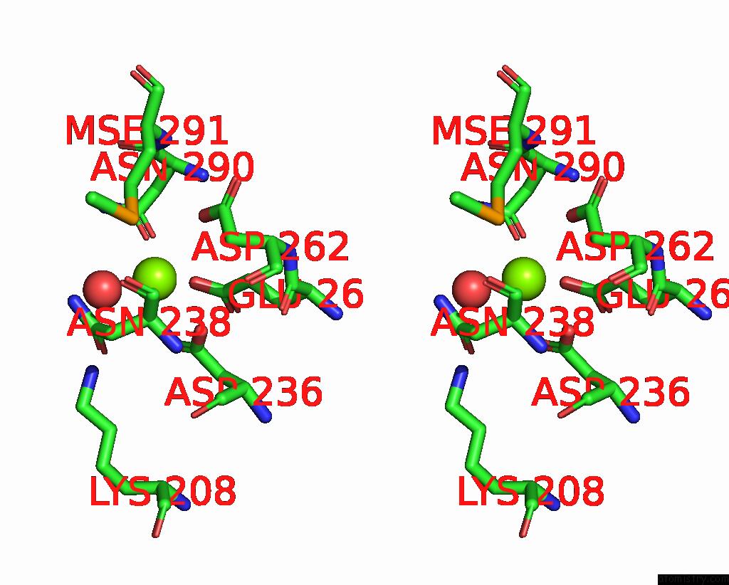
Stereo pair view
Mono view
Stereo pair view
A full contact list of Magnesium with other atoms in the Mg binding
site number 2 of Crystal Structure of Mandelate Racemase/Muconate Lactonizing Protein From Actinobacillus Succinogenes 130Z Complexed with Magnesium/Sulfate within 5.0Å range:
|
Magnesium binding site 3 out of 4 in 3n6h
Go back to
Magnesium binding site 3 out
of 4 in the Crystal Structure of Mandelate Racemase/Muconate Lactonizing Protein From Actinobacillus Succinogenes 130Z Complexed with Magnesium/Sulfate
Mono view
Stereo pair view
Mono view
Stereo pair view
A full contact list of Magnesium with other atoms in the Mg binding
site number 3 of Crystal Structure of Mandelate Racemase/Muconate Lactonizing Protein From Actinobacillus Succinogenes 130Z Complexed with Magnesium/Sulfate within 5.0Å range:
|
Magnesium binding site 4 out of 4 in 3n6h
Go back to
Magnesium binding site 4 out
of 4 in the Crystal Structure of Mandelate Racemase/Muconate Lactonizing Protein From Actinobacillus Succinogenes 130Z Complexed with Magnesium/Sulfate
Mono view
Stereo pair view
Mono view
Stereo pair view
A full contact list of Magnesium with other atoms in the Mg binding
site number 4 of Crystal Structure of Mandelate Racemase/Muconate Lactonizing Protein From Actinobacillus Succinogenes 130Z Complexed with Magnesium/Sulfate within 5.0Å range:
|
Reference:
V.N.Malashkevich,
R.Toro,
J.M.Sauder,
S.K.Burley,
S.C.Almo.
Crystal Structure of Mandelate Racemase/Muconate Lactonizing Protein From Actinobacillus Succinogenes 130Z Complexed with Magnesium/Sulfate To Be Published.
Page generated: Thu Aug 15 07:51:55 2024
Last articles
Ca in 5TF9Ca in 5TAK
Ca in 5TAE
Ca in 5TAJ
Ca in 5TAI
Ca in 5T9T
Ca in 5TAD
Ca in 5T5O
Ca in 5TA5
Ca in 5TAC