Magnesium »
PDB 3n8u-3nky »
3na7 »
Magnesium in PDB 3na7: 2.2 Angstrom Structure of the HP0958 Protein From Helicobacter Pylori Ccug 17874
Protein crystallography data
The structure of 2.2 Angstrom Structure of the HP0958 Protein From Helicobacter Pylori Ccug 17874, PDB code: 3na7
was solved by
D.L.Caly,
P.W.O'toole,
S.A.Moore,
with X-Ray Crystallography technique. A brief refinement statistics is given in the table below:
| Resolution Low / High (Å) | 30.00 / 2.20 |
| Space group | C 1 2 1 |
| Cell size a, b, c (Å), α, β, γ (°) | 172.720, 37.951, 66.056, 90.00, 110.66, 90.00 |
| R / Rfree (%) | 24.8 / 29.4 |
Other elements in 3na7:
The structure of 2.2 Angstrom Structure of the HP0958 Protein From Helicobacter Pylori Ccug 17874 also contains other interesting chemical elements:
| Zinc | (Zn) | 1 atom |
Magnesium Binding Sites:
The binding sites of Magnesium atom in the 2.2 Angstrom Structure of the HP0958 Protein From Helicobacter Pylori Ccug 17874
(pdb code 3na7). This binding sites where shown within
5.0 Angstroms radius around Magnesium atom.
In total only one binding site of Magnesium was determined in the 2.2 Angstrom Structure of the HP0958 Protein From Helicobacter Pylori Ccug 17874, PDB code: 3na7:
In total only one binding site of Magnesium was determined in the 2.2 Angstrom Structure of the HP0958 Protein From Helicobacter Pylori Ccug 17874, PDB code: 3na7:
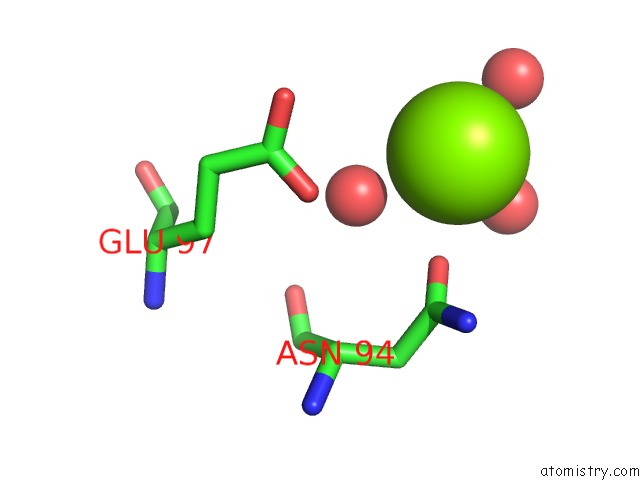
Magnesium binding site 1 out of 1 in 3na7
Go back to
Magnesium binding site 1 out
of 1 in the 2.2 Angstrom Structure of the HP0958 Protein From Helicobacter Pylori Ccug 17874
Mono view

Stereo pair view
Mono view
Stereo pair view
A full contact list of Magnesium with other atoms in the Mg binding
site number 1 of 2.2 Angstrom Structure of the HP0958 Protein From Helicobacter Pylori Ccug 17874 within 5.0Å range:
|
Reference:
D.L.Caly,
P.W.O'toole,
S.A.Moore.
The 2.2-A Structure of the HP0958 Protein From Helicobacter Pylori Reveals A Kinked Anti-Parallel Coiled-Coil Hairpin Domain and A Highly Conserved Zn-Ribbon Domain J.Mol.Biol. V. 403 405 2010.
ISSN: ISSN 0022-2836
PubMed: 20826163
DOI: 10.1016/J.JMB.2010.08.051
Page generated: Thu Aug 15 07:55:38 2024
ISSN: ISSN 0022-2836
PubMed: 20826163
DOI: 10.1016/J.JMB.2010.08.051
Last articles
Fe in 2YXOFe in 2YRS
Fe in 2YXC
Fe in 2YNM
Fe in 2YVJ
Fe in 2YP1
Fe in 2YU2
Fe in 2YU1
Fe in 2YQB
Fe in 2YOO