Magnesium »
PDB 3n8u-3nky »
3ngq »
Magnesium in PDB 3ngq: Crystal Structure of the Human CNOT6L Nuclease Domain
Protein crystallography data
The structure of Crystal Structure of the Human CNOT6L Nuclease Domain, PDB code: 3ngq
was solved by
H.Wang,
M.Morita,
W.Yang,
M.Bartlam,
T.Yamamoto,
Z.Rao,
with X-Ray Crystallography technique. A brief refinement statistics is given in the table below:
| Resolution Low / High (Å) | 50.00 / 1.80 |
| Space group | P 32 2 1 |
| Cell size a, b, c (Å), α, β, γ (°) | 77.152, 77.152, 165.897, 90.00, 90.00, 120.00 |
| R / Rfree (%) | 22 / 25.4 |
Magnesium Binding Sites:
The binding sites of Magnesium atom in the Crystal Structure of the Human CNOT6L Nuclease Domain
(pdb code 3ngq). This binding sites where shown within
5.0 Angstroms radius around Magnesium atom.
In total 2 binding sites of Magnesium where determined in the Crystal Structure of the Human CNOT6L Nuclease Domain, PDB code: 3ngq:
Jump to Magnesium binding site number: 1; 2;
In total 2 binding sites of Magnesium where determined in the Crystal Structure of the Human CNOT6L Nuclease Domain, PDB code: 3ngq:
Jump to Magnesium binding site number: 1; 2;
Magnesium binding site 1 out of 2 in 3ngq
Go back to
Magnesium binding site 1 out
of 2 in the Crystal Structure of the Human CNOT6L Nuclease Domain
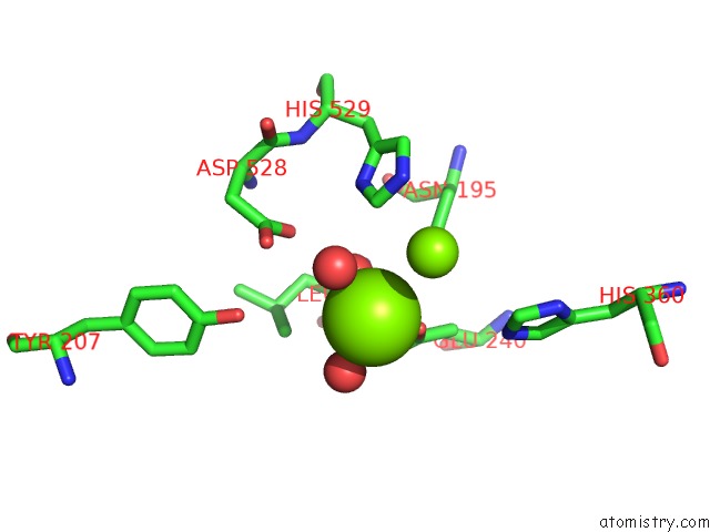
Mono view
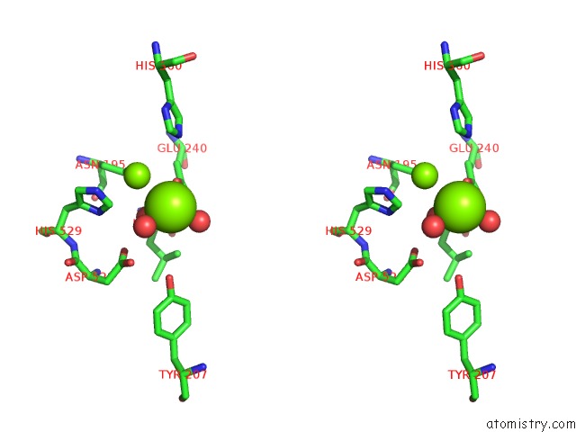
Stereo pair view
Mono view
Stereo pair view
A full contact list of Magnesium with other atoms in the Mg binding
site number 1 of Crystal Structure of the Human CNOT6L Nuclease Domain within 5.0Å range:
|
Magnesium binding site 2 out of 2 in 3ngq
Go back to
Magnesium binding site 2 out
of 2 in the Crystal Structure of the Human CNOT6L Nuclease Domain
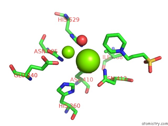
Mono view
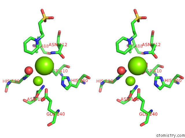
Stereo pair view
Mono view
Stereo pair view
A full contact list of Magnesium with other atoms in the Mg binding
site number 2 of Crystal Structure of the Human CNOT6L Nuclease Domain within 5.0Å range:
|
Reference:
H.Wang,
M.Morita,
X.Yang,
T.Suzuki,
W.Yang,
J.Wang,
K.Ito,
Q.Wang,
C.Zhao,
M.Bartlam,
T.Yamamoto,
Z.Rao.
Crystal Structure of the Human CNOT6L Nuclease Domain Reveals Strict Poly(A) Substrate Specificity. Embo J. 2010.
ISSN: ESSN 1460-2075
PubMed: 20628353
DOI: 10.1038/EMBOJ.2010.152
Page generated: Mon Aug 11 00:53:49 2025
ISSN: ESSN 1460-2075
PubMed: 20628353
DOI: 10.1038/EMBOJ.2010.152
Last articles
Mg in 6PRVMg in 6PSS
Mg in 6PSR
Mg in 6PSQ
Mg in 6PRY
Mg in 6PRU
Mg in 6PRC
Mg in 6PR5
Mg in 6PQV
Mg in 6PQR