Magnesium »
PDB 3nl0-3o0q »
3no3 »
Magnesium in PDB 3no3: Crystal Structure of A Glycerophosphodiester Phosphodiesterase (BDI_0402) From Parabacteroides Distasonis Atcc 8503 at 1.89 A Resolution
Protein crystallography data
The structure of Crystal Structure of A Glycerophosphodiester Phosphodiesterase (BDI_0402) From Parabacteroides Distasonis Atcc 8503 at 1.89 A Resolution, PDB code: 3no3
was solved by
Joint Center For Structural Genomics (Jcsg),
with X-Ray Crystallography technique. A brief refinement statistics is given in the table below:
| Resolution Low / High (Å) | 26.95 / 1.89 |
| Space group | C 1 2 1 |
| Cell size a, b, c (Å), α, β, γ (°) | 137.460, 40.876, 48.328, 90.00, 99.39, 90.00 |
| R / Rfree (%) | 15 / 17.7 |
Magnesium Binding Sites:
The binding sites of Magnesium atom in the Crystal Structure of A Glycerophosphodiester Phosphodiesterase (BDI_0402) From Parabacteroides Distasonis Atcc 8503 at 1.89 A Resolution
(pdb code 3no3). This binding sites where shown within
5.0 Angstroms radius around Magnesium atom.
In total 2 binding sites of Magnesium where determined in the Crystal Structure of A Glycerophosphodiester Phosphodiesterase (BDI_0402) From Parabacteroides Distasonis Atcc 8503 at 1.89 A Resolution, PDB code: 3no3:
Jump to Magnesium binding site number: 1; 2;
In total 2 binding sites of Magnesium where determined in the Crystal Structure of A Glycerophosphodiester Phosphodiesterase (BDI_0402) From Parabacteroides Distasonis Atcc 8503 at 1.89 A Resolution, PDB code: 3no3:
Jump to Magnesium binding site number: 1; 2;
Magnesium binding site 1 out of 2 in 3no3
Go back to
Magnesium binding site 1 out
of 2 in the Crystal Structure of A Glycerophosphodiester Phosphodiesterase (BDI_0402) From Parabacteroides Distasonis Atcc 8503 at 1.89 A Resolution
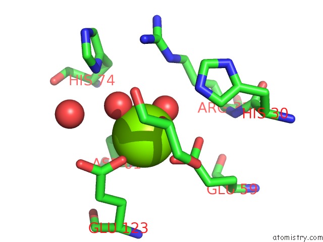
Mono view
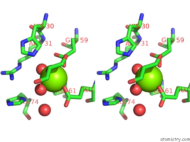
Stereo pair view
Mono view
Stereo pair view
A full contact list of Magnesium with other atoms in the Mg binding
site number 1 of Crystal Structure of A Glycerophosphodiester Phosphodiesterase (BDI_0402) From Parabacteroides Distasonis Atcc 8503 at 1.89 A Resolution within 5.0Å range:
|
Magnesium binding site 2 out of 2 in 3no3
Go back to
Magnesium binding site 2 out
of 2 in the Crystal Structure of A Glycerophosphodiester Phosphodiesterase (BDI_0402) From Parabacteroides Distasonis Atcc 8503 at 1.89 A Resolution
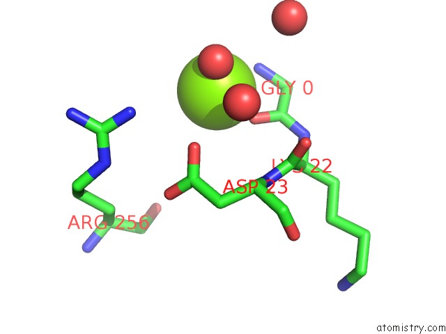
Mono view
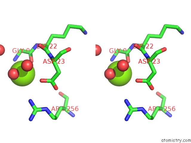
Stereo pair view
Mono view
Stereo pair view
A full contact list of Magnesium with other atoms in the Mg binding
site number 2 of Crystal Structure of A Glycerophosphodiester Phosphodiesterase (BDI_0402) From Parabacteroides Distasonis Atcc 8503 at 1.89 A Resolution within 5.0Å range:
|
Reference:
Joint Center For Structural Genomics (Jcsg),
Joint Center For Structural Genomics (Jcsg).
N/A N/A.
Page generated: Thu Aug 15 08:05:53 2024
Last articles
Cl in 7VSLCl in 7VSI
Cl in 7VQM
Cl in 7VR9
Cl in 7VPE
Cl in 7VRE
Cl in 7VRA
Cl in 7VP8
Cl in 7VOE
Cl in 7VOD