Magnesium »
PDB 3o1n-3oha »
3o69 »
Magnesium in PDB 3o69: Structure of the E100A E.Coli Gdp-Mannose Hydrolase (Yffh) in Complex with Mg++
Protein crystallography data
The structure of Structure of the E100A E.Coli Gdp-Mannose Hydrolase (Yffh) in Complex with Mg++, PDB code: 3o69
was solved by
L.M.Amzel,
S.B.Gabelli,
A.N.Boto,
with X-Ray Crystallography technique. A brief refinement statistics is given in the table below:
| Resolution Low / High (Å) | 22.81 / 2.10 |
| Space group | P 21 21 21 |
| Cell size a, b, c (Å), α, β, γ (°) | 60.379, 69.260, 98.554, 90.00, 90.00, 90.00 |
| R / Rfree (%) | 20.5 / 28 |
Other elements in 3o69:
The structure of Structure of the E100A E.Coli Gdp-Mannose Hydrolase (Yffh) in Complex with Mg++ also contains other interesting chemical elements:
| Chlorine | (Cl) | 3 atoms |
| Sodium | (Na) | 3 atoms |
Magnesium Binding Sites:
The binding sites of Magnesium atom in the Structure of the E100A E.Coli Gdp-Mannose Hydrolase (Yffh) in Complex with Mg++
(pdb code 3o69). This binding sites where shown within
5.0 Angstroms radius around Magnesium atom.
In total 4 binding sites of Magnesium where determined in the Structure of the E100A E.Coli Gdp-Mannose Hydrolase (Yffh) in Complex with Mg++, PDB code: 3o69:
Jump to Magnesium binding site number: 1; 2; 3; 4;
In total 4 binding sites of Magnesium where determined in the Structure of the E100A E.Coli Gdp-Mannose Hydrolase (Yffh) in Complex with Mg++, PDB code: 3o69:
Jump to Magnesium binding site number: 1; 2; 3; 4;
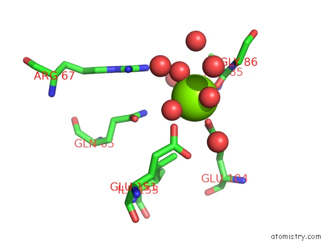
Magnesium binding site 1 out of 4 in 3o69
Go back to
Magnesium binding site 1 out
of 4 in the Structure of the E100A E.Coli Gdp-Mannose Hydrolase (Yffh) in Complex with Mg++
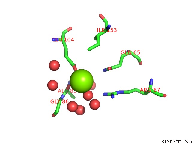
Mono view
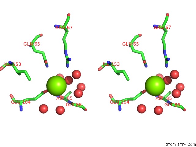
Stereo pair view
Mono view
Stereo pair view
A full contact list of Magnesium with other atoms in the Mg binding
site number 1 of Structure of the E100A E.Coli Gdp-Mannose Hydrolase (Yffh) in Complex with Mg++ within 5.0Å range:
|
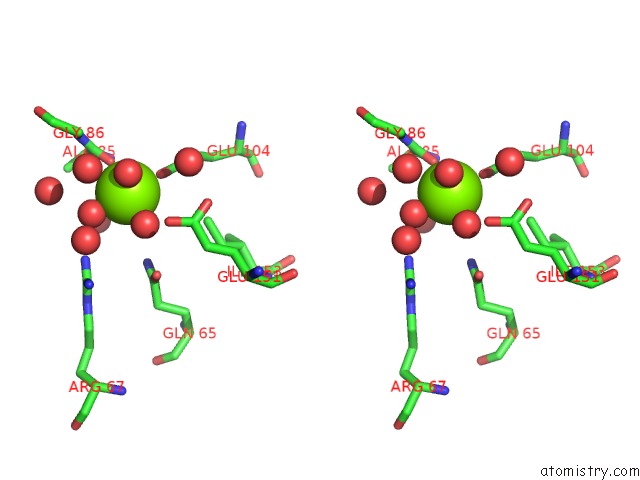
Magnesium binding site 2 out of 4 in 3o69
Go back to
Magnesium binding site 2 out
of 4 in the Structure of the E100A E.Coli Gdp-Mannose Hydrolase (Yffh) in Complex with Mg++
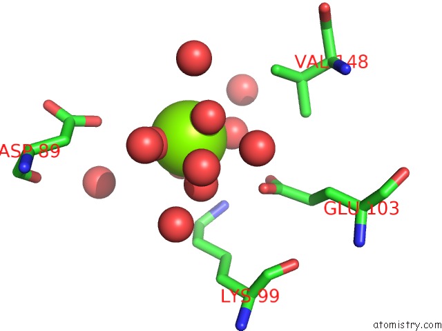
Mono view
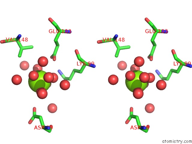
Stereo pair view
Mono view
Stereo pair view
A full contact list of Magnesium with other atoms in the Mg binding
site number 2 of Structure of the E100A E.Coli Gdp-Mannose Hydrolase (Yffh) in Complex with Mg++ within 5.0Å range:
|
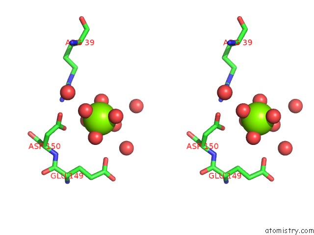
Magnesium binding site 3 out of 4 in 3o69
Go back to
Magnesium binding site 3 out
of 4 in the Structure of the E100A E.Coli Gdp-Mannose Hydrolase (Yffh) in Complex with Mg++
Mono view
Stereo pair view
Mono view
Stereo pair view
A full contact list of Magnesium with other atoms in the Mg binding
site number 3 of Structure of the E100A E.Coli Gdp-Mannose Hydrolase (Yffh) in Complex with Mg++ within 5.0Å range:
|
Magnesium binding site 4 out of 4 in 3o69
Go back to
Magnesium binding site 4 out
of 4 in the Structure of the E100A E.Coli Gdp-Mannose Hydrolase (Yffh) in Complex with Mg++

Mono view
Stereo pair view
Mono view
Stereo pair view
A full contact list of Magnesium with other atoms in the Mg binding
site number 4 of Structure of the E100A E.Coli Gdp-Mannose Hydrolase (Yffh) in Complex with Mg++ within 5.0Å range:
|
Reference:
A.N.Boto,
W.Xu,
J.Jakoncic,
A.Pannuri,
T.Romeo,
M.J.Bessman,
S.B.Gabelli,
L.M.Amzel.
Structural Studies of the Nudix Gdp-Mannose Hydrolase From E. Coli Reveals A New Motif For Mannose Recognition. Proteins V. 79 2455 2011.
ISSN: ISSN 0887-3585
PubMed: 21638333
DOI: 10.1002/PROT.23069
Page generated: Thu Aug 15 08:12:03 2024
ISSN: ISSN 0887-3585
PubMed: 21638333
DOI: 10.1002/PROT.23069
Last articles
Zn in 9J0NZn in 9J0O
Zn in 9J0P
Zn in 9FJX
Zn in 9EKB
Zn in 9C0F
Zn in 9CAH
Zn in 9CH0
Zn in 9CH3
Zn in 9CH1