Magnesium »
PDB 3o1n-3oha »
3ogh »
Magnesium in PDB 3ogh: Crystal Structure of Ycie Protein From E. Coli CFT073, A Member of Ferritine-Like Superfamily of Diiron-Containing Four-Helix-Bundle Proteins
Protein crystallography data
The structure of Crystal Structure of Ycie Protein From E. Coli CFT073, A Member of Ferritine-Like Superfamily of Diiron-Containing Four-Helix-Bundle Proteins, PDB code: 3ogh
was solved by
B.Nocek,
L.Bigelow,
J.Bearden,
A.Joachimiak,
Midwest Center Forstructural Genomics (Mcsg),
with X-Ray Crystallography technique. A brief refinement statistics is given in the table below:
| Resolution Low / High (Å) | 40.00 / 1.65 |
| Space group | P 1 21 1 |
| Cell size a, b, c (Å), α, β, γ (°) | 30.148, 90.295, 58.132, 90.00, 103.51, 90.00 |
| R / Rfree (%) | 16.6 / 21.3 |
Other elements in 3ogh:
The structure of Crystal Structure of Ycie Protein From E. Coli CFT073, A Member of Ferritine-Like Superfamily of Diiron-Containing Four-Helix-Bundle Proteins also contains other interesting chemical elements:
| Iron | (Fe) | 2 atoms |
| Chlorine | (Cl) | 1 atom |
Magnesium Binding Sites:
The binding sites of Magnesium atom in the Crystal Structure of Ycie Protein From E. Coli CFT073, A Member of Ferritine-Like Superfamily of Diiron-Containing Four-Helix-Bundle Proteins
(pdb code 3ogh). This binding sites where shown within
5.0 Angstroms radius around Magnesium atom.
In total 2 binding sites of Magnesium where determined in the Crystal Structure of Ycie Protein From E. Coli CFT073, A Member of Ferritine-Like Superfamily of Diiron-Containing Four-Helix-Bundle Proteins, PDB code: 3ogh:
Jump to Magnesium binding site number: 1; 2;
In total 2 binding sites of Magnesium where determined in the Crystal Structure of Ycie Protein From E. Coli CFT073, A Member of Ferritine-Like Superfamily of Diiron-Containing Four-Helix-Bundle Proteins, PDB code: 3ogh:
Jump to Magnesium binding site number: 1; 2;
Magnesium binding site 1 out of 2 in 3ogh
Go back to
Magnesium binding site 1 out
of 2 in the Crystal Structure of Ycie Protein From E. Coli CFT073, A Member of Ferritine-Like Superfamily of Diiron-Containing Four-Helix-Bundle Proteins
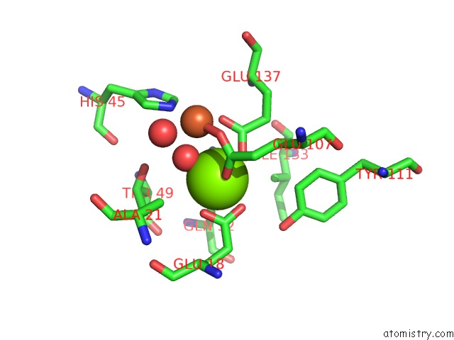
Mono view
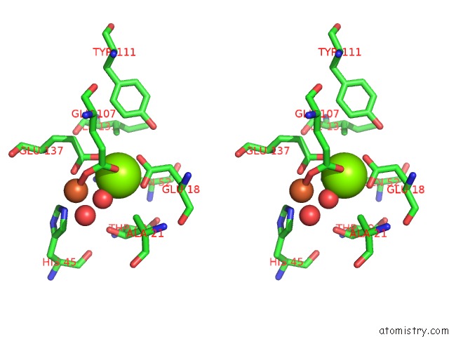
Stereo pair view
Mono view
Stereo pair view
A full contact list of Magnesium with other atoms in the Mg binding
site number 1 of Crystal Structure of Ycie Protein From E. Coli CFT073, A Member of Ferritine-Like Superfamily of Diiron-Containing Four-Helix-Bundle Proteins within 5.0Å range:
|
Magnesium binding site 2 out of 2 in 3ogh
Go back to
Magnesium binding site 2 out
of 2 in the Crystal Structure of Ycie Protein From E. Coli CFT073, A Member of Ferritine-Like Superfamily of Diiron-Containing Four-Helix-Bundle Proteins
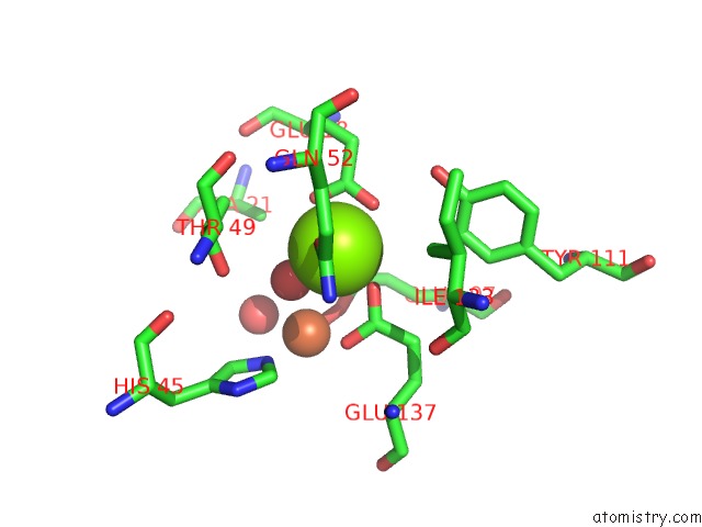
Mono view

Stereo pair view
Mono view
Stereo pair view
A full contact list of Magnesium with other atoms in the Mg binding
site number 2 of Crystal Structure of Ycie Protein From E. Coli CFT073, A Member of Ferritine-Like Superfamily of Diiron-Containing Four-Helix-Bundle Proteins within 5.0Å range:
|
Reference:
B.Nocek,
L.Bigelow,
J.Bearden,
A.Joachimiak.
Crystal Structure of Ycie Protein From E. Coli CFT073, A Member of Ferritine-Like Superfamily of Diiron-Containing Four-Helix-Bundle Proteins To Be Published.
Page generated: Thu Aug 15 08:23:05 2024
Last articles
Zn in 9MJ5Zn in 9HNW
Zn in 9G0L
Zn in 9FNE
Zn in 9DZN
Zn in 9E0I
Zn in 9D32
Zn in 9DAK
Zn in 8ZXC
Zn in 8ZUF