Magnesium »
PDB 3ohb-3otb »
3osu »
Magnesium in PDB 3osu: Crystal Structure of the 3-Oxoacyl-Acyl Carrier Protein Reductase, Fabg, From Staphylococcus Aureus
Enzymatic activity of Crystal Structure of the 3-Oxoacyl-Acyl Carrier Protein Reductase, Fabg, From Staphylococcus Aureus
All present enzymatic activity of Crystal Structure of the 3-Oxoacyl-Acyl Carrier Protein Reductase, Fabg, From Staphylococcus Aureus:
1.1.1.100;
1.1.1.100;
Protein crystallography data
The structure of Crystal Structure of the 3-Oxoacyl-Acyl Carrier Protein Reductase, Fabg, From Staphylococcus Aureus, PDB code: 3osu
was solved by
S.M.Anderson,
Z.Wawrzak,
O.Onopriyenko,
A.Edwards,
W.F.Anderson,
A.Savchenko,
Center For Structural Genomics Of Infectious Diseases(Csgid),
with X-Ray Crystallography technique. A brief refinement statistics is given in the table below:
| Resolution Low / High (Å) | 32.03 / 1.90 |
| Space group | P 31 2 1 |
| Cell size a, b, c (Å), α, β, γ (°) | 64.060, 64.060, 183.890, 90.00, 90.00, 120.00 |
| R / Rfree (%) | 15.7 / 19.3 |
Magnesium Binding Sites:
The binding sites of Magnesium atom in the Crystal Structure of the 3-Oxoacyl-Acyl Carrier Protein Reductase, Fabg, From Staphylococcus Aureus
(pdb code 3osu). This binding sites where shown within
5.0 Angstroms radius around Magnesium atom.
In total 2 binding sites of Magnesium where determined in the Crystal Structure of the 3-Oxoacyl-Acyl Carrier Protein Reductase, Fabg, From Staphylococcus Aureus, PDB code: 3osu:
Jump to Magnesium binding site number: 1; 2;
In total 2 binding sites of Magnesium where determined in the Crystal Structure of the 3-Oxoacyl-Acyl Carrier Protein Reductase, Fabg, From Staphylococcus Aureus, PDB code: 3osu:
Jump to Magnesium binding site number: 1; 2;
Magnesium binding site 1 out of 2 in 3osu
Go back to
Magnesium binding site 1 out
of 2 in the Crystal Structure of the 3-Oxoacyl-Acyl Carrier Protein Reductase, Fabg, From Staphylococcus Aureus
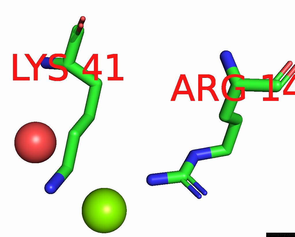
Mono view
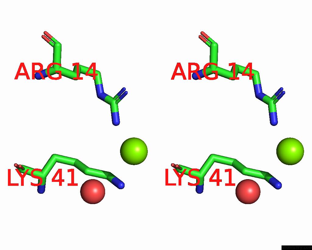
Stereo pair view
Mono view
Stereo pair view
A full contact list of Magnesium with other atoms in the Mg binding
site number 1 of Crystal Structure of the 3-Oxoacyl-Acyl Carrier Protein Reductase, Fabg, From Staphylococcus Aureus within 5.0Å range:
|
Magnesium binding site 2 out of 2 in 3osu
Go back to
Magnesium binding site 2 out
of 2 in the Crystal Structure of the 3-Oxoacyl-Acyl Carrier Protein Reductase, Fabg, From Staphylococcus Aureus
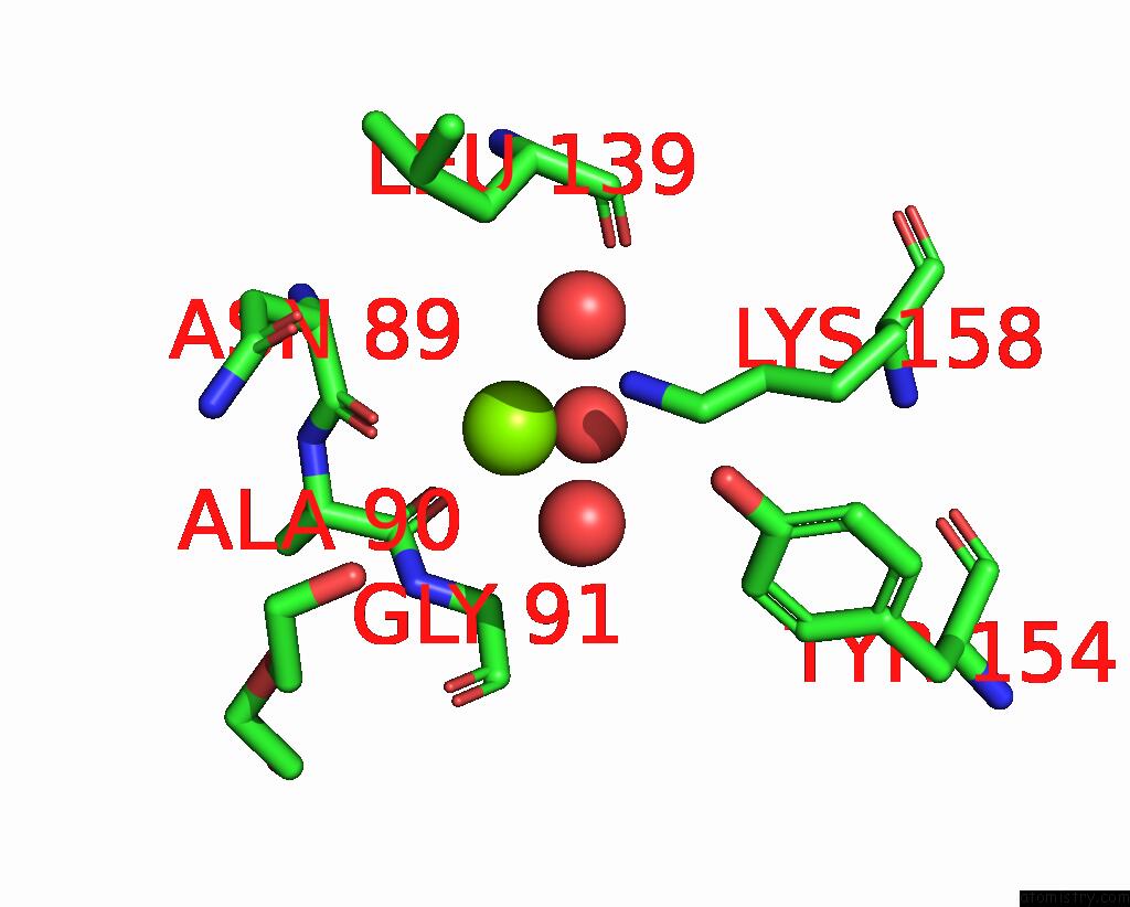
Mono view

Stereo pair view
Mono view
Stereo pair view
A full contact list of Magnesium with other atoms in the Mg binding
site number 2 of Crystal Structure of the 3-Oxoacyl-Acyl Carrier Protein Reductase, Fabg, From Staphylococcus Aureus within 5.0Å range:
|
Reference:
S.M.Anderson,
Z.Wawrzak,
O.Onopriyenko,
A.Edwards,
W.F.Anderson,
A.Savchenko.
Crystal Structure of the 3-Oxoacyl-Acyl Carrier Protein Reductase, Fabg, From Staphylococcus Aureus To Be Published.
Page generated: Mon Aug 11 01:17:09 2025
Last articles
Mg in 4APZMg in 4AV6
Mg in 4AVA
Mg in 4AV3
Mg in 4AUX
Mg in 4ATB
Mg in 4AUI
Mg in 4AT9
Mg in 4AT8
Mg in 4AS5