Magnesium »
PDB 3oto-3p0x »
3ow1 »
Magnesium in PDB 3ow1: Crystal Structure of D-Mannonate Dehydratase From Chromohalobacter Salexigens Complexed with Mg
Protein crystallography data
The structure of Crystal Structure of D-Mannonate Dehydratase From Chromohalobacter Salexigens Complexed with Mg, PDB code: 3ow1
was solved by
A.A.Fedorov,
E.V.Fedorov,
D.Wichelecki,
J.A.Gerlt,
S.C.Almo,
with X-Ray Crystallography technique. A brief refinement statistics is given in the table below:
| Resolution Low / High (Å) | 35.17 / 1.80 |
| Space group | P 1 21 1 |
| Cell size a, b, c (Å), α, β, γ (°) | 86.585, 177.787, 109.993, 90.00, 102.62, 90.00 |
| R / Rfree (%) | 16.1 / 19.4 |
Magnesium Binding Sites:
The binding sites of Magnesium atom in the Crystal Structure of D-Mannonate Dehydratase From Chromohalobacter Salexigens Complexed with Mg
(pdb code 3ow1). This binding sites where shown within
5.0 Angstroms radius around Magnesium atom.
In total 8 binding sites of Magnesium where determined in the Crystal Structure of D-Mannonate Dehydratase From Chromohalobacter Salexigens Complexed with Mg, PDB code: 3ow1:
Jump to Magnesium binding site number: 1; 2; 3; 4; 5; 6; 7; 8;
In total 8 binding sites of Magnesium where determined in the Crystal Structure of D-Mannonate Dehydratase From Chromohalobacter Salexigens Complexed with Mg, PDB code: 3ow1:
Jump to Magnesium binding site number: 1; 2; 3; 4; 5; 6; 7; 8;
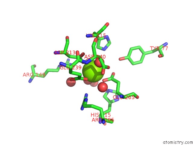
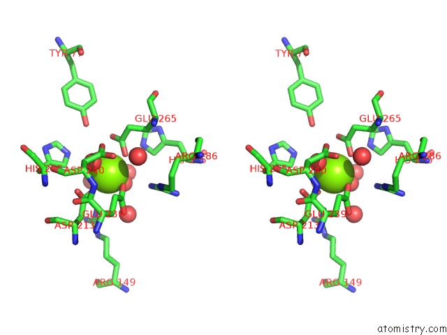
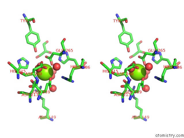
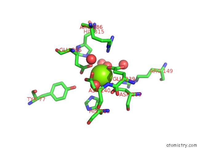
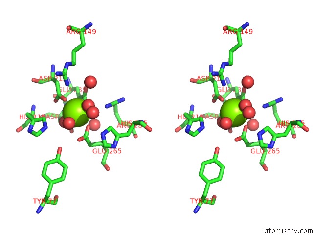
Magnesium binding site 1 out of 8 in 3ow1
Go back to
Magnesium binding site 1 out
of 8 in the Crystal Structure of D-Mannonate Dehydratase From Chromohalobacter Salexigens Complexed with Mg
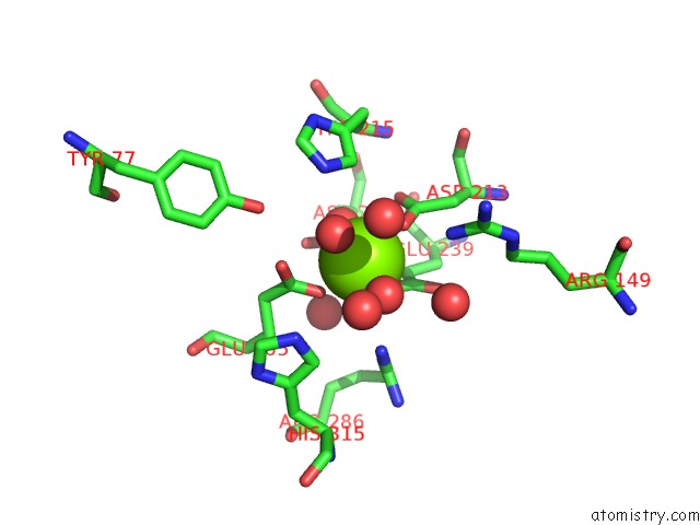
Mono view
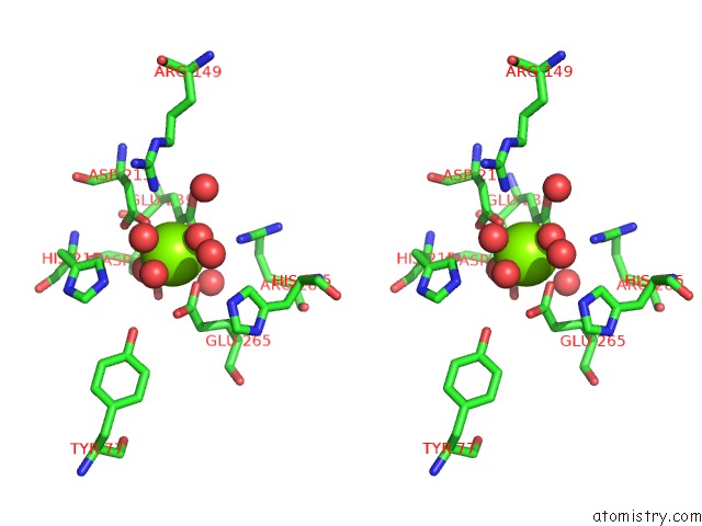
Stereo pair view
Mono view
Stereo pair view
A full contact list of Magnesium with other atoms in the Mg binding
site number 1 of Crystal Structure of D-Mannonate Dehydratase From Chromohalobacter Salexigens Complexed with Mg within 5.0Å range:
|
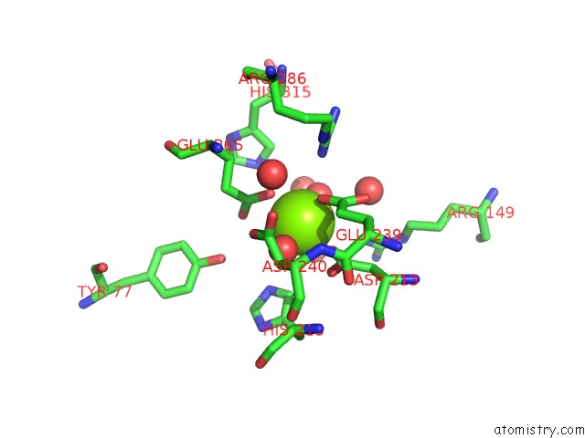
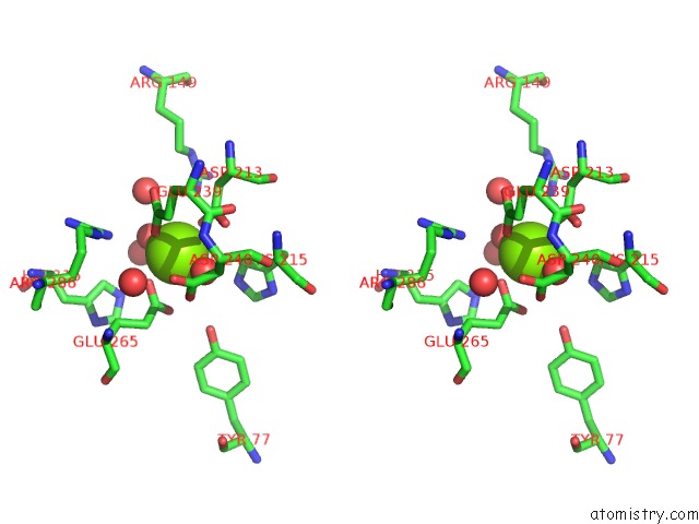
Magnesium binding site 2 out of 8 in 3ow1
Go back to
Magnesium binding site 2 out
of 8 in the Crystal Structure of D-Mannonate Dehydratase From Chromohalobacter Salexigens Complexed with Mg
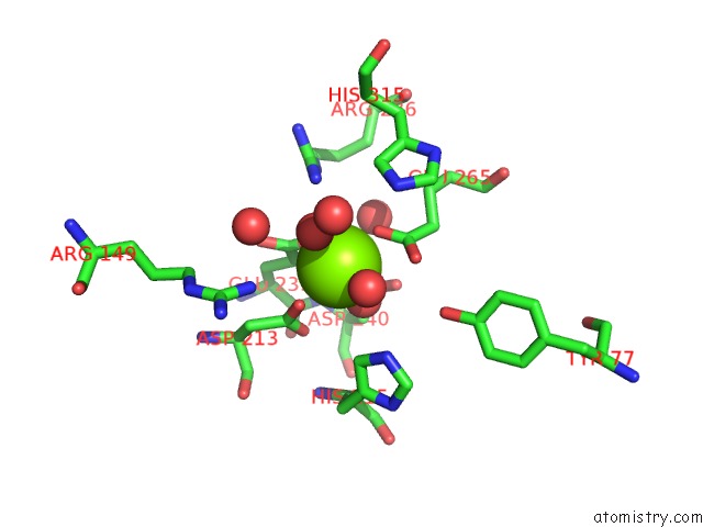
Mono view
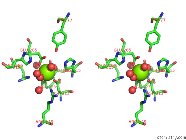
Stereo pair view
Mono view
Stereo pair view
A full contact list of Magnesium with other atoms in the Mg binding
site number 2 of Crystal Structure of D-Mannonate Dehydratase From Chromohalobacter Salexigens Complexed with Mg within 5.0Å range:
|
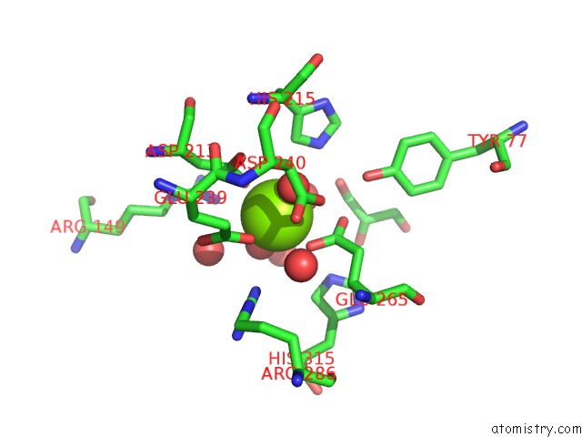
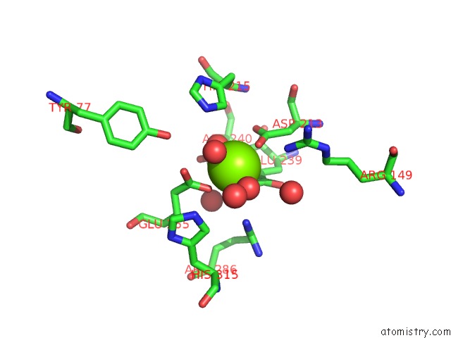
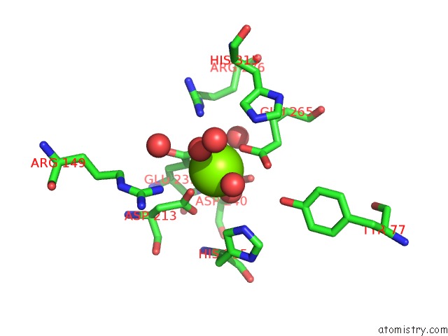
Magnesium binding site 3 out of 8 in 3ow1
Go back to
Magnesium binding site 3 out
of 8 in the Crystal Structure of D-Mannonate Dehydratase From Chromohalobacter Salexigens Complexed with Mg
Mono view
Stereo pair view
Mono view
Stereo pair view
A full contact list of Magnesium with other atoms in the Mg binding
site number 3 of Crystal Structure of D-Mannonate Dehydratase From Chromohalobacter Salexigens Complexed with Mg within 5.0Å range:
|
Magnesium binding site 4 out of 8 in 3ow1
Go back to
Magnesium binding site 4 out
of 8 in the Crystal Structure of D-Mannonate Dehydratase From Chromohalobacter Salexigens Complexed with Mg
Mono view

Stereo pair view
Mono view
Stereo pair view
A full contact list of Magnesium with other atoms in the Mg binding
site number 4 of Crystal Structure of D-Mannonate Dehydratase From Chromohalobacter Salexigens Complexed with Mg within 5.0Å range:
|
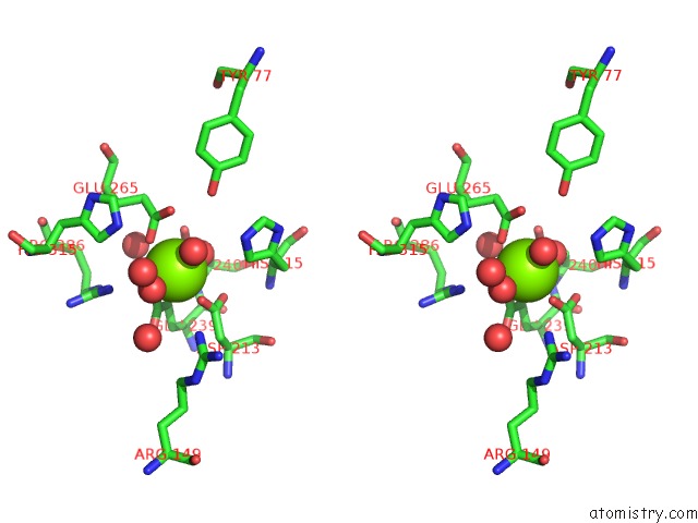
Magnesium binding site 5 out of 8 in 3ow1
Go back to
Magnesium binding site 5 out
of 8 in the Crystal Structure of D-Mannonate Dehydratase From Chromohalobacter Salexigens Complexed with Mg
Mono view
Stereo pair view
Mono view
Stereo pair view
A full contact list of Magnesium with other atoms in the Mg binding
site number 5 of Crystal Structure of D-Mannonate Dehydratase From Chromohalobacter Salexigens Complexed with Mg within 5.0Å range:
|
Magnesium binding site 6 out of 8 in 3ow1
Go back to
Magnesium binding site 6 out
of 8 in the Crystal Structure of D-Mannonate Dehydratase From Chromohalobacter Salexigens Complexed with Mg
Mono view
Stereo pair view
Mono view
Stereo pair view
A full contact list of Magnesium with other atoms in the Mg binding
site number 6 of Crystal Structure of D-Mannonate Dehydratase From Chromohalobacter Salexigens Complexed with Mg within 5.0Å range:
|
Magnesium binding site 7 out of 8 in 3ow1
Go back to
Magnesium binding site 7 out
of 8 in the Crystal Structure of D-Mannonate Dehydratase From Chromohalobacter Salexigens Complexed with Mg
Mono view
Stereo pair view
Mono view
Stereo pair view
A full contact list of Magnesium with other atoms in the Mg binding
site number 7 of Crystal Structure of D-Mannonate Dehydratase From Chromohalobacter Salexigens Complexed with Mg within 5.0Å range:
|
Magnesium binding site 8 out of 8 in 3ow1
Go back to
Magnesium binding site 8 out
of 8 in the Crystal Structure of D-Mannonate Dehydratase From Chromohalobacter Salexigens Complexed with Mg
Mono view
Stereo pair view
Mono view
Stereo pair view
A full contact list of Magnesium with other atoms in the Mg binding
site number 8 of Crystal Structure of D-Mannonate Dehydratase From Chromohalobacter Salexigens Complexed with Mg within 5.0Å range:
|
Reference:
A.A.Fedorov,
E.V.Fedorov,
D.Wichelecki,
J.A.Gerlt,
S.C.Almo.
Crystal Structure of D-Mannonate Dehydratase From Chromohalobacter Salexigens Complexed with Mg To Be Published.
Page generated: Mon Aug 11 01:18:05 2025
Last articles
Mg in 3TZ5Mg in 3TZ4
Mg in 3TXA
Mg in 3TY7
Mg in 3TX9
Mg in 3TXZ
Mg in 3TWP
Mg in 3TWB
Mg in 3TWH
Mg in 3TW6