Magnesium »
PDB 3oto-3p0x »
3oww »
Magnesium in PDB 3oww: Crystal Structure of the Glycine Riboswitch Bound to Glycine
Protein crystallography data
The structure of Crystal Structure of the Glycine Riboswitch Bound to Glycine, PDB code: 3oww
was solved by
L.Huang,
A.Serganov,
D.J.Patel,
with X-Ray Crystallography technique. A brief refinement statistics is given in the table below:
| Resolution Low / High (Å) | 20.00 / 2.80 |
| Space group | P 32 2 1 |
| Cell size a, b, c (Å), α, β, γ (°) | 83.514, 83.514, 198.524, 90.00, 90.00, 120.00 |
| R / Rfree (%) | 20.5 / 23.6 |
Magnesium Binding Sites:
Pages:
>>> Page 1 <<< Page 2, Binding sites: 11 - 20; Page 3, Binding sites: 21 - 30; Page 4, Binding sites: 31 - 33;Binding sites:
The binding sites of Magnesium atom in the Crystal Structure of the Glycine Riboswitch Bound to Glycine (pdb code 3oww). This binding sites where shown within 5.0 Angstroms radius around Magnesium atom.In total 33 binding sites of Magnesium where determined in the Crystal Structure of the Glycine Riboswitch Bound to Glycine, PDB code: 3oww:
Jump to Magnesium binding site number: 1; 2; 3; 4; 5; 6; 7; 8; 9; 10;
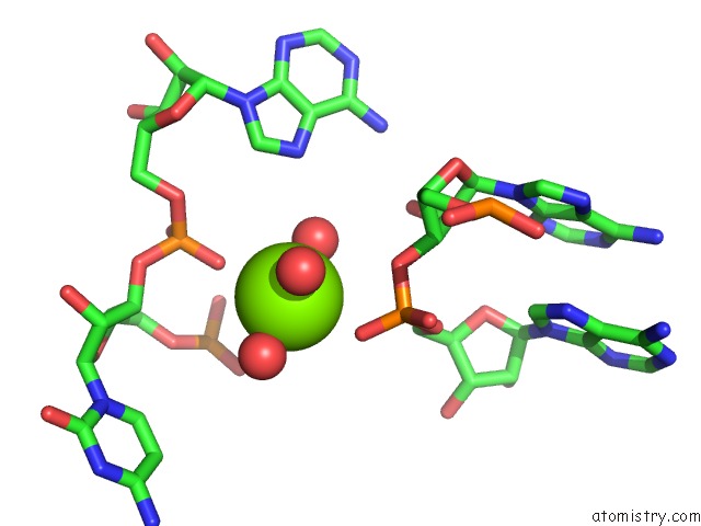
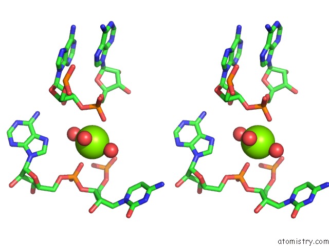
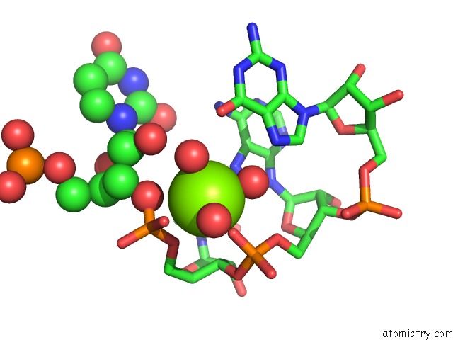
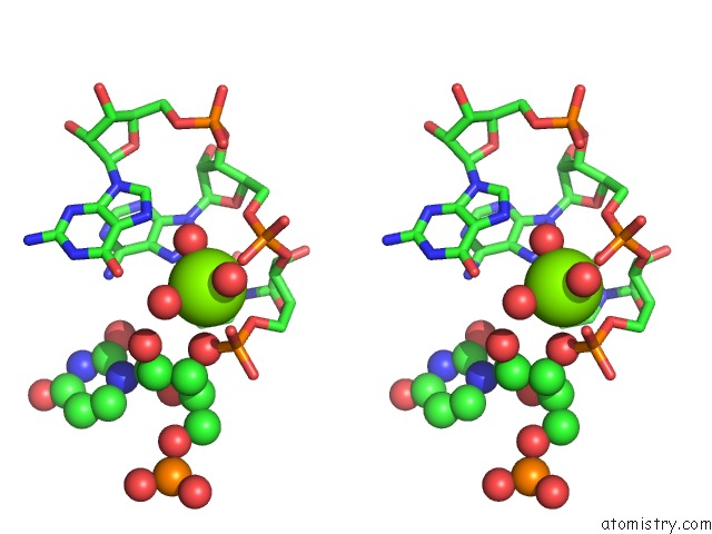
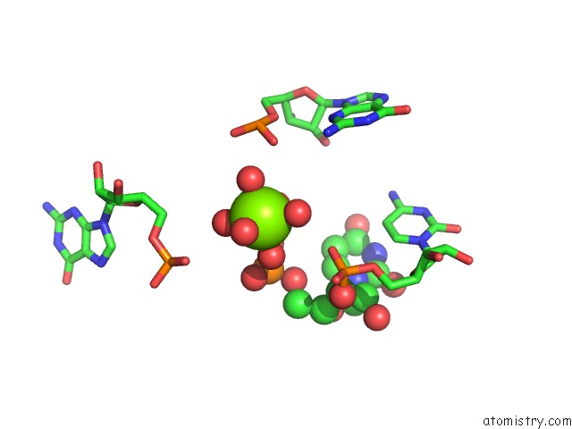
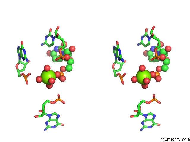
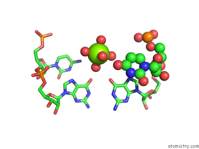
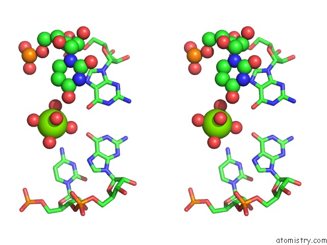
Magnesium binding site 1 out of 33 in 3oww
Go back to
Magnesium binding site 1 out
of 33 in the Crystal Structure of the Glycine Riboswitch Bound to Glycine
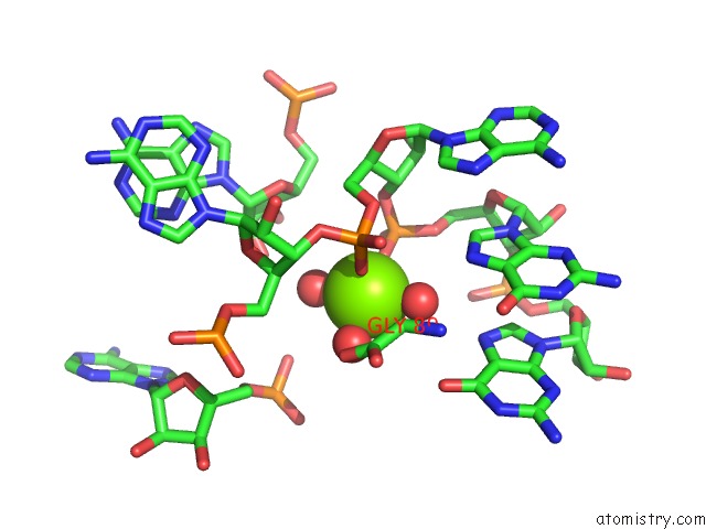
Mono view
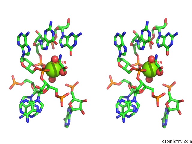
Stereo pair view
Mono view
Stereo pair view
A full contact list of Magnesium with other atoms in the Mg binding
site number 1 of Crystal Structure of the Glycine Riboswitch Bound to Glycine within 5.0Å range:
|
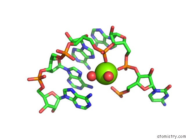
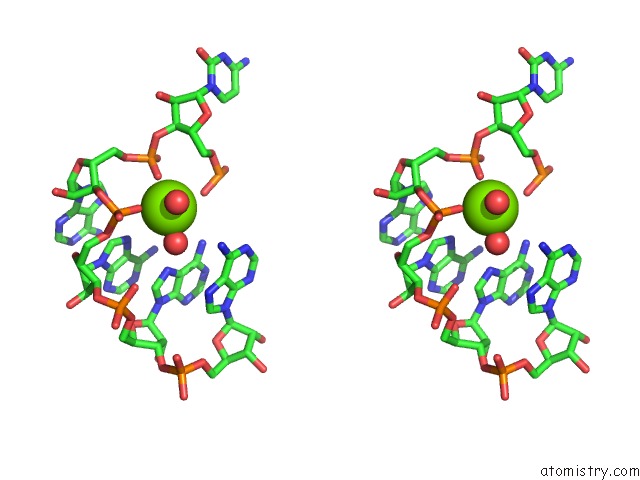
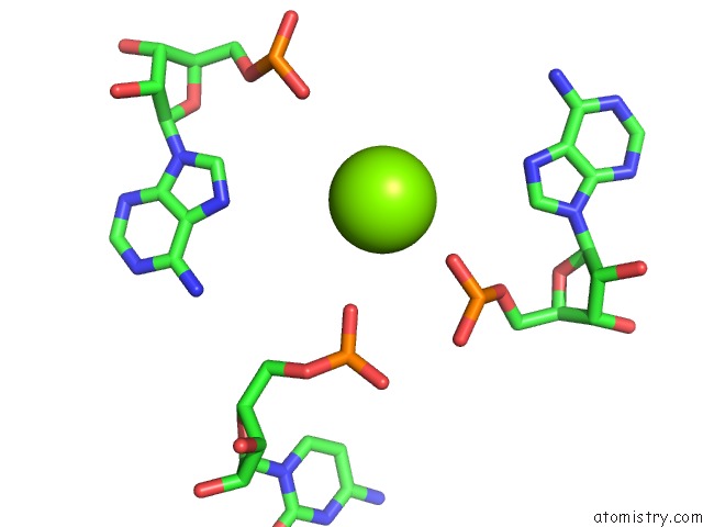
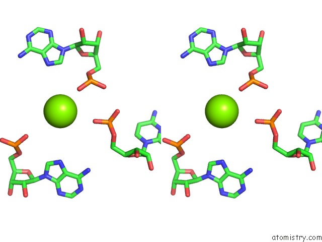
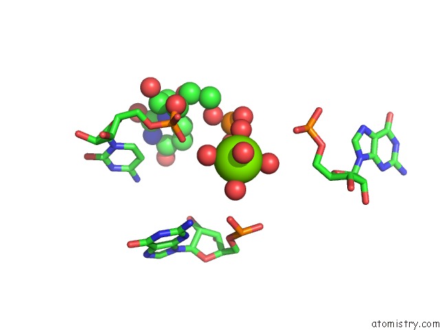
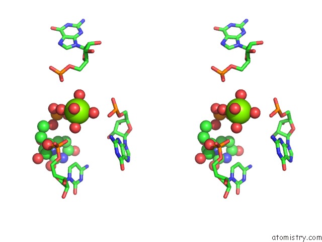
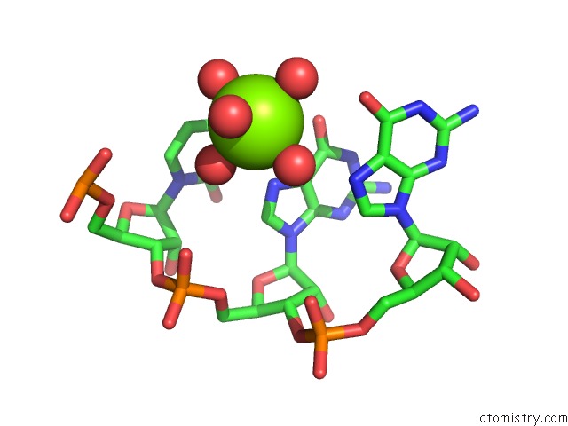
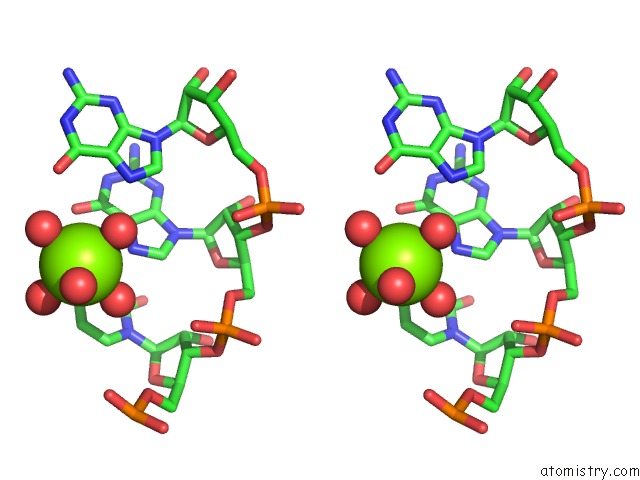
Magnesium binding site 2 out of 33 in 3oww
Go back to
Magnesium binding site 2 out
of 33 in the Crystal Structure of the Glycine Riboswitch Bound to Glycine
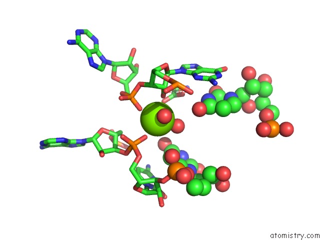
Mono view
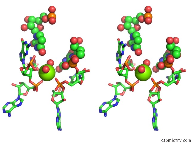
Stereo pair view
Mono view
Stereo pair view
A full contact list of Magnesium with other atoms in the Mg binding
site number 2 of Crystal Structure of the Glycine Riboswitch Bound to Glycine within 5.0Å range:
|
Magnesium binding site 3 out of 33 in 3oww
Go back to
Magnesium binding site 3 out
of 33 in the Crystal Structure of the Glycine Riboswitch Bound to Glycine
Mono view
Stereo pair view
Mono view
Stereo pair view
A full contact list of Magnesium with other atoms in the Mg binding
site number 3 of Crystal Structure of the Glycine Riboswitch Bound to Glycine within 5.0Å range:
|
Magnesium binding site 4 out of 33 in 3oww
Go back to
Magnesium binding site 4 out
of 33 in the Crystal Structure of the Glycine Riboswitch Bound to Glycine
Mono view
Stereo pair view
Mono view
Stereo pair view
A full contact list of Magnesium with other atoms in the Mg binding
site number 4 of Crystal Structure of the Glycine Riboswitch Bound to Glycine within 5.0Å range:
|
Magnesium binding site 5 out of 33 in 3oww
Go back to
Magnesium binding site 5 out
of 33 in the Crystal Structure of the Glycine Riboswitch Bound to Glycine
Mono view
Stereo pair view
Mono view
Stereo pair view
A full contact list of Magnesium with other atoms in the Mg binding
site number 5 of Crystal Structure of the Glycine Riboswitch Bound to Glycine within 5.0Å range:
|
Magnesium binding site 6 out of 33 in 3oww
Go back to
Magnesium binding site 6 out
of 33 in the Crystal Structure of the Glycine Riboswitch Bound to Glycine
Mono view
Stereo pair view
Mono view
Stereo pair view
A full contact list of Magnesium with other atoms in the Mg binding
site number 6 of Crystal Structure of the Glycine Riboswitch Bound to Glycine within 5.0Å range:
|
Magnesium binding site 7 out of 33 in 3oww
Go back to
Magnesium binding site 7 out
of 33 in the Crystal Structure of the Glycine Riboswitch Bound to Glycine
Mono view
Stereo pair view
Mono view
Stereo pair view
A full contact list of Magnesium with other atoms in the Mg binding
site number 7 of Crystal Structure of the Glycine Riboswitch Bound to Glycine within 5.0Å range:
|
Magnesium binding site 8 out of 33 in 3oww
Go back to
Magnesium binding site 8 out
of 33 in the Crystal Structure of the Glycine Riboswitch Bound to Glycine
Mono view
Stereo pair view
Mono view
Stereo pair view
A full contact list of Magnesium with other atoms in the Mg binding
site number 8 of Crystal Structure of the Glycine Riboswitch Bound to Glycine within 5.0Å range:
|
Magnesium binding site 9 out of 33 in 3oww
Go back to
Magnesium binding site 9 out
of 33 in the Crystal Structure of the Glycine Riboswitch Bound to Glycine
Mono view
Stereo pair view
Mono view
Stereo pair view
A full contact list of Magnesium with other atoms in the Mg binding
site number 9 of Crystal Structure of the Glycine Riboswitch Bound to Glycine within 5.0Å range:
|
Magnesium binding site 10 out of 33 in 3oww
Go back to
Magnesium binding site 10 out
of 33 in the Crystal Structure of the Glycine Riboswitch Bound to Glycine
Mono view
Stereo pair view
Mono view
Stereo pair view
A full contact list of Magnesium with other atoms in the Mg binding
site number 10 of Crystal Structure of the Glycine Riboswitch Bound to Glycine within 5.0Å range:
|
Reference:
L.Huang,
A.Serganov,
D.J.Patel.
Structural Insights Into Ligand Recognition By A Sensing Domain of the Cooperative Glycine Riboswitch. Mol.Cell V. 40 774 2010.
ISSN: ISSN 1097-2765
PubMed: 21145485
DOI: 10.1016/J.MOLCEL.2010.11.026
Page generated: Mon Aug 11 01:20:18 2025
ISSN: ISSN 1097-2765
PubMed: 21145485
DOI: 10.1016/J.MOLCEL.2010.11.026
Last articles
Mg in 4HN2Mg in 4HNS
Mg in 4HNQ
Mg in 4HNO
Mg in 4HNA
Mg in 4HNL
Mg in 4HNC
Mg in 4HMY
Mg in 4HLQ
Mg in 4HHM