Magnesium »
PDB 3oto-3p0x »
3oya »
Magnesium in PDB 3oya: Crystal Structure of the Prototype Foamy Virus (Pfv) Intasome in Complex with Magnesium and Raltegravir at 2.65 Resolution
Protein crystallography data
The structure of Crystal Structure of the Prototype Foamy Virus (Pfv) Intasome in Complex with Magnesium and Raltegravir at 2.65 Resolution, PDB code: 3oya
was solved by
S.Hare,
P.Cherepanov,
with X-Ray Crystallography technique. A brief refinement statistics is given in the table below:
| Resolution Low / High (Å) | 38.02 / 2.65 |
| Space group | P 41 21 2 |
| Cell size a, b, c (Å), α, β, γ (°) | 159.755, 159.755, 124.139, 90.00, 90.00, 90.00 |
| R / Rfree (%) | 20.5 / 22.8 |
Other elements in 3oya:
The structure of Crystal Structure of the Prototype Foamy Virus (Pfv) Intasome in Complex with Magnesium and Raltegravir at 2.65 Resolution also contains other interesting chemical elements:
| Fluorine | (F) | 1 atom |
| Zinc | (Zn) | 1 atom |
Magnesium Binding Sites:
The binding sites of Magnesium atom in the Crystal Structure of the Prototype Foamy Virus (Pfv) Intasome in Complex with Magnesium and Raltegravir at 2.65 Resolution
(pdb code 3oya). This binding sites where shown within
5.0 Angstroms radius around Magnesium atom.
In total 3 binding sites of Magnesium where determined in the Crystal Structure of the Prototype Foamy Virus (Pfv) Intasome in Complex with Magnesium and Raltegravir at 2.65 Resolution, PDB code: 3oya:
Jump to Magnesium binding site number: 1; 2; 3;
In total 3 binding sites of Magnesium where determined in the Crystal Structure of the Prototype Foamy Virus (Pfv) Intasome in Complex with Magnesium and Raltegravir at 2.65 Resolution, PDB code: 3oya:
Jump to Magnesium binding site number: 1; 2; 3;
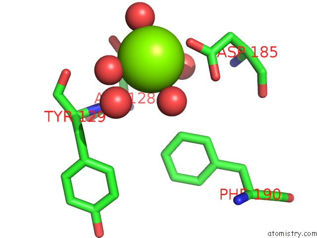
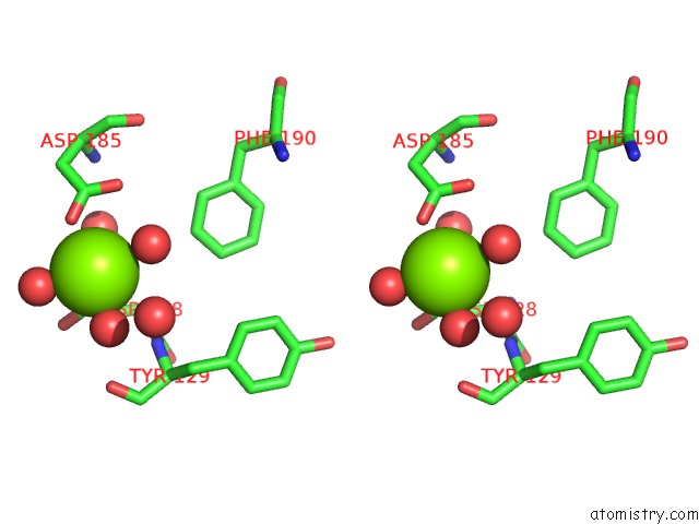
Magnesium binding site 1 out of 3 in 3oya
Go back to
Magnesium binding site 1 out
of 3 in the Crystal Structure of the Prototype Foamy Virus (Pfv) Intasome in Complex with Magnesium and Raltegravir at 2.65 Resolution
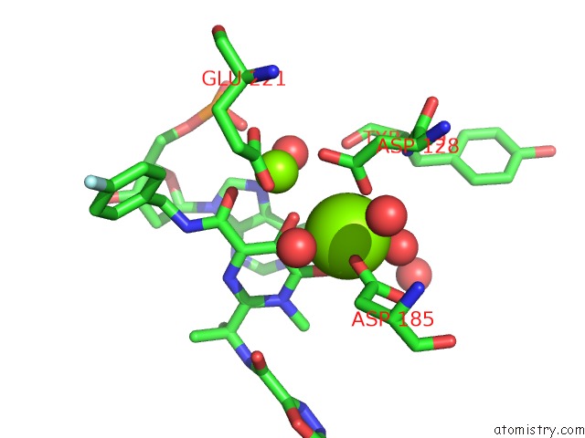
Mono view
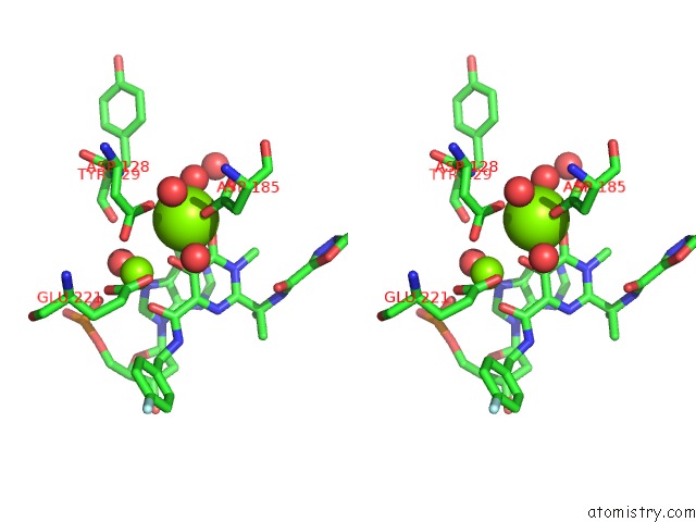
Stereo pair view
Mono view
Stereo pair view
A full contact list of Magnesium with other atoms in the Mg binding
site number 1 of Crystal Structure of the Prototype Foamy Virus (Pfv) Intasome in Complex with Magnesium and Raltegravir at 2.65 Resolution within 5.0Å range:
|
Magnesium binding site 2 out of 3 in 3oya
Go back to
Magnesium binding site 2 out
of 3 in the Crystal Structure of the Prototype Foamy Virus (Pfv) Intasome in Complex with Magnesium and Raltegravir at 2.65 Resolution
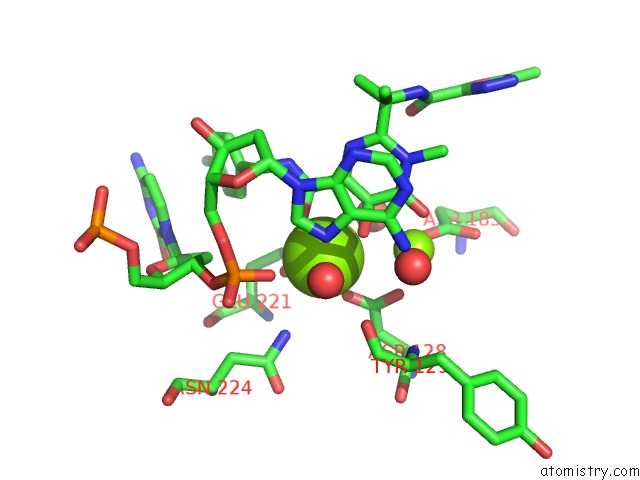
Mono view
Stereo pair view
Mono view
Stereo pair view
A full contact list of Magnesium with other atoms in the Mg binding
site number 2 of Crystal Structure of the Prototype Foamy Virus (Pfv) Intasome in Complex with Magnesium and Raltegravir at 2.65 Resolution within 5.0Å range:
|
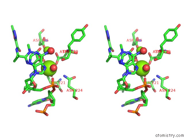
Magnesium binding site 3 out of 3 in 3oya
Go back to
Magnesium binding site 3 out
of 3 in the Crystal Structure of the Prototype Foamy Virus (Pfv) Intasome in Complex with Magnesium and Raltegravir at 2.65 Resolution
Mono view
Stereo pair view
Mono view
Stereo pair view
A full contact list of Magnesium with other atoms in the Mg binding
site number 3 of Crystal Structure of the Prototype Foamy Virus (Pfv) Intasome in Complex with Magnesium and Raltegravir at 2.65 Resolution within 5.0Å range:
|
Reference:
S.Hare,
A.M.Vos,
R.F.Clayton,
J.W.Thuring,
M.D.Cummings,
P.Cherepanov.
Molecular Mechanisms of Retroviral Integrase Inhibition and the Evolution of Viral Resistance. Proc.Natl.Acad.Sci.Usa V. 107 20057 2010.
ISSN: ISSN 0027-8424
PubMed: 21030679
DOI: 10.1073/PNAS.1010246107
Page generated: Thu Aug 15 08:51:06 2024
ISSN: ISSN 0027-8424
PubMed: 21030679
DOI: 10.1073/PNAS.1010246107
Last articles
Zn in 9J0NZn in 9J0O
Zn in 9J0P
Zn in 9FJX
Zn in 9EKB
Zn in 9C0F
Zn in 9CAH
Zn in 9CH0
Zn in 9CH3
Zn in 9CH1