Magnesium »
PDB 3oto-3p0x »
3p0f »
Magnesium in PDB 3p0f: Structure of HUPP2 in An Inactive Conformation with Bound 5- Benzylacyclouridine
Enzymatic activity of Structure of HUPP2 in An Inactive Conformation with Bound 5- Benzylacyclouridine
All present enzymatic activity of Structure of HUPP2 in An Inactive Conformation with Bound 5- Benzylacyclouridine:
2.4.2.3;
2.4.2.3;
Protein crystallography data
The structure of Structure of HUPP2 in An Inactive Conformation with Bound 5- Benzylacyclouridine, PDB code: 3p0f
was solved by
T.P.Roosild,
S.Castronovo,
A.Villoso,
with X-Ray Crystallography technique. A brief refinement statistics is given in the table below:
| Resolution Low / High (Å) | 50.00 / 1.54 |
| Space group | P 43 21 2 |
| Cell size a, b, c (Å), α, β, γ (°) | 59.696, 59.696, 189.307, 90.00, 90.00, 90.00 |
| R / Rfree (%) | 19.1 / 21.2 |
Other elements in 3p0f:
The structure of Structure of HUPP2 in An Inactive Conformation with Bound 5- Benzylacyclouridine also contains other interesting chemical elements:
| Cobalt | (Co) | 2 atoms |
Magnesium Binding Sites:
The binding sites of Magnesium atom in the Structure of HUPP2 in An Inactive Conformation with Bound 5- Benzylacyclouridine
(pdb code 3p0f). This binding sites where shown within
5.0 Angstroms radius around Magnesium atom.
In total only one binding site of Magnesium was determined in the Structure of HUPP2 in An Inactive Conformation with Bound 5- Benzylacyclouridine, PDB code: 3p0f:
In total only one binding site of Magnesium was determined in the Structure of HUPP2 in An Inactive Conformation with Bound 5- Benzylacyclouridine, PDB code: 3p0f:
Magnesium binding site 1 out of 1 in 3p0f
Go back to
Magnesium binding site 1 out
of 1 in the Structure of HUPP2 in An Inactive Conformation with Bound 5- Benzylacyclouridine
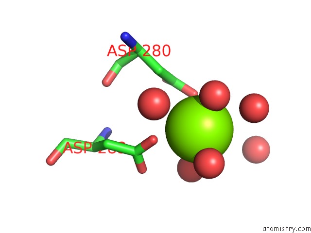
Mono view
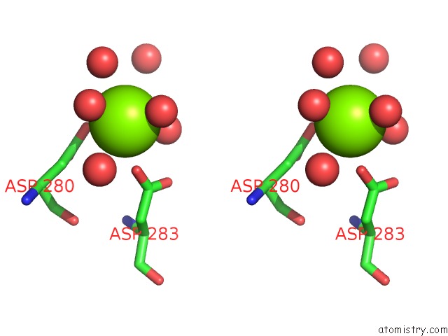
Stereo pair view
Mono view
Stereo pair view
A full contact list of Magnesium with other atoms in the Mg binding
site number 1 of Structure of HUPP2 in An Inactive Conformation with Bound 5- Benzylacyclouridine within 5.0Å range:
|
Reference:
T.P.Roosild,
S.Castronovo,
A.Villoso,
A.Ziemba,
G.Pizzorno.
A Novel Structural Mechanism For Redox Regulation of Uridine Phosphorylase 2 Activity. J.Struct.Biol. V. 176 229 2011.
ISSN: ISSN 1047-8477
PubMed: 21855639
DOI: 10.1016/J.JSB.2011.08.002
Page generated: Thu Aug 15 09:01:41 2024
ISSN: ISSN 1047-8477
PubMed: 21855639
DOI: 10.1016/J.JSB.2011.08.002
Last articles
Zn in 9J0NZn in 9J0O
Zn in 9J0P
Zn in 9FJX
Zn in 9EKB
Zn in 9C0F
Zn in 9CAH
Zn in 9CH0
Zn in 9CH3
Zn in 9CH1