Magnesium »
PDB 3p1g-3pio »
3p1g »
Magnesium in PDB 3p1g: Crystal Structure of the Xenotropic Murine Leukemia Virus-Related Virus (Xmrv) Rnase H Domain
Protein crystallography data
The structure of Crystal Structure of the Xenotropic Murine Leukemia Virus-Related Virus (Xmrv) Rnase H Domain, PDB code: 3p1g
was solved by
K.A.Kirby,
S.G.Sarafianos,
with X-Ray Crystallography technique. A brief refinement statistics is given in the table below:
| Resolution Low / High (Å) | 37.53 / 1.50 |
| Space group | P 41 |
| Cell size a, b, c (Å), α, β, γ (°) | 37.530, 37.530, 100.720, 90.00, 90.00, 90.00 |
| R / Rfree (%) | 16.1 / 18.2 |
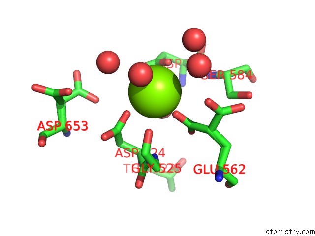
Magnesium Binding Sites:
The binding sites of Magnesium atom in the Crystal Structure of the Xenotropic Murine Leukemia Virus-Related Virus (Xmrv) Rnase H Domain
(pdb code 3p1g). This binding sites where shown within
5.0 Angstroms radius around Magnesium atom.
In total only one binding site of Magnesium was determined in the Crystal Structure of the Xenotropic Murine Leukemia Virus-Related Virus (Xmrv) Rnase H Domain, PDB code: 3p1g:
In total only one binding site of Magnesium was determined in the Crystal Structure of the Xenotropic Murine Leukemia Virus-Related Virus (Xmrv) Rnase H Domain, PDB code: 3p1g:
Magnesium binding site 1 out of 1 in 3p1g
Go back to
Magnesium binding site 1 out
of 1 in the Crystal Structure of the Xenotropic Murine Leukemia Virus-Related Virus (Xmrv) Rnase H Domain
Mono view

Stereo pair view
Mono view
Stereo pair view
A full contact list of Magnesium with other atoms in the Mg binding
site number 1 of Crystal Structure of the Xenotropic Murine Leukemia Virus-Related Virus (Xmrv) Rnase H Domain within 5.0Å range:
|
Reference:
K.A.Kirby,
B.Marchand,
Y.T.Ong,
T.P.Ndongwe,
A.Hachiya,
E.Michailidis,
M.D.Leslie,
D.V.Sietsema,
T.L.Fetterly,
C.A.Dorst,
K.Singh,
Z.Wang,
M.A.Parniak,
S.G.Sarafianos.
Structural and Inhibition Studies of the Rnase H Function of Xenotropic Murine Leukemia Virus-Related Virus Reverse Transcriptase. Antimicrob.Agents Chemother. V. 56 2048 2012.
ISSN: ISSN 0066-4804
PubMed: 22252812
DOI: 10.1128/AAC.06000-11
Page generated: Thu Aug 15 09:05:42 2024
ISSN: ISSN 0066-4804
PubMed: 22252812
DOI: 10.1128/AAC.06000-11
Last articles
F in 4J0BF in 4IYN
F in 4J0T
F in 4J03
F in 4J0P
F in 4IZW
F in 4IW8
F in 4IZT
F in 4IWF
F in 4IXE