Magnesium »
PDB 3p1g-3pio »
3pc8 »
Magnesium in PDB 3pc8: X-Ray Crystal Structure of the Heterodimeric Complex of XRCC1 and Dna Ligase III-Alpha Brct Domains.
Enzymatic activity of X-Ray Crystal Structure of the Heterodimeric Complex of XRCC1 and Dna Ligase III-Alpha Brct Domains.
All present enzymatic activity of X-Ray Crystal Structure of the Heterodimeric Complex of XRCC1 and Dna Ligase III-Alpha Brct Domains.:
6.5.1.1;
6.5.1.1;
Protein crystallography data
The structure of X-Ray Crystal Structure of the Heterodimeric Complex of XRCC1 and Dna Ligase III-Alpha Brct Domains., PDB code: 3pc8
was solved by
M.J.Cuneo,
J.M.Krahn,
R.E.London,
with X-Ray Crystallography technique. A brief refinement statistics is given in the table below:
| Resolution Low / High (Å) | 33.51 / 2.31 |
| Space group | P 21 21 2 |
| Cell size a, b, c (Å), α, β, γ (°) | 66.320, 163.480, 36.740, 90.00, 90.00, 90.00 |
| R / Rfree (%) | 19.7 / 24.6 |
Magnesium Binding Sites:
The binding sites of Magnesium atom in the X-Ray Crystal Structure of the Heterodimeric Complex of XRCC1 and Dna Ligase III-Alpha Brct Domains.
(pdb code 3pc8). This binding sites where shown within
5.0 Angstroms radius around Magnesium atom.
In total only one binding site of Magnesium was determined in the X-Ray Crystal Structure of the Heterodimeric Complex of XRCC1 and Dna Ligase III-Alpha Brct Domains., PDB code: 3pc8:
In total only one binding site of Magnesium was determined in the X-Ray Crystal Structure of the Heterodimeric Complex of XRCC1 and Dna Ligase III-Alpha Brct Domains., PDB code: 3pc8:
Magnesium binding site 1 out of 1 in 3pc8
Go back to
Magnesium binding site 1 out
of 1 in the X-Ray Crystal Structure of the Heterodimeric Complex of XRCC1 and Dna Ligase III-Alpha Brct Domains.
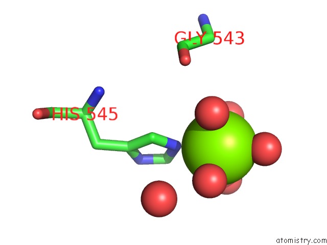
Mono view
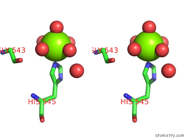
Stereo pair view
Mono view
Stereo pair view
A full contact list of Magnesium with other atoms in the Mg binding
site number 1 of X-Ray Crystal Structure of the Heterodimeric Complex of XRCC1 and Dna Ligase III-Alpha Brct Domains. within 5.0Å range:
|
Reference:
M.J.Cuneo,
S.A.Gabel,
J.M.Krahn,
M.A.Ricker,
R.E.London.
The Structural Basis For Partitioning of the XRCC1/Dna Ligase III-{Alpha} Brct-Mediated Dimer Complexes. Nucleic Acids Res. V. 39 7816 2011.
ISSN: ISSN 0305-1048
PubMed: 21652643
DOI: 10.1093/NAR/GKR419
Page generated: Thu Aug 15 09:10:04 2024
ISSN: ISSN 0305-1048
PubMed: 21652643
DOI: 10.1093/NAR/GKR419
Last articles
Fe in 2YXOFe in 2YRS
Fe in 2YXC
Fe in 2YNM
Fe in 2YVJ
Fe in 2YP1
Fe in 2YU2
Fe in 2YU1
Fe in 2YQB
Fe in 2YOO