Magnesium »
PDB 3p1g-3pio »
3pdt »
Magnesium in PDB 3pdt: Crystal Structure of the C-Terminal Truncated Alpha-Kinase Domain of Myosin Heavy Chain Kinase
Enzymatic activity of Crystal Structure of the C-Terminal Truncated Alpha-Kinase Domain of Myosin Heavy Chain Kinase
All present enzymatic activity of Crystal Structure of the C-Terminal Truncated Alpha-Kinase Domain of Myosin Heavy Chain Kinase:
2.7.11.7;
2.7.11.7;
Protein crystallography data
The structure of Crystal Structure of the C-Terminal Truncated Alpha-Kinase Domain of Myosin Heavy Chain Kinase, PDB code: 3pdt
was solved by
Q.Ye,
Z.Jia,
with X-Ray Crystallography technique. A brief refinement statistics is given in the table below:
| Resolution Low / High (Å) | 28.39 / 1.80 |
| Space group | P 21 21 2 |
| Cell size a, b, c (Å), α, β, γ (°) | 77.062, 83.681, 44.684, 90.00, 90.00, 90.00 |
| R / Rfree (%) | 19.9 / 24.3 |
Other elements in 3pdt:
The structure of Crystal Structure of the C-Terminal Truncated Alpha-Kinase Domain of Myosin Heavy Chain Kinase also contains other interesting chemical elements:
| Zinc | (Zn) | 1 atom |
Magnesium Binding Sites:
The binding sites of Magnesium atom in the Crystal Structure of the C-Terminal Truncated Alpha-Kinase Domain of Myosin Heavy Chain Kinase
(pdb code 3pdt). This binding sites where shown within
5.0 Angstroms radius around Magnesium atom.
In total 2 binding sites of Magnesium where determined in the Crystal Structure of the C-Terminal Truncated Alpha-Kinase Domain of Myosin Heavy Chain Kinase, PDB code: 3pdt:
Jump to Magnesium binding site number: 1; 2;
In total 2 binding sites of Magnesium where determined in the Crystal Structure of the C-Terminal Truncated Alpha-Kinase Domain of Myosin Heavy Chain Kinase, PDB code: 3pdt:
Jump to Magnesium binding site number: 1; 2;
Magnesium binding site 1 out of 2 in 3pdt
Go back to
Magnesium binding site 1 out
of 2 in the Crystal Structure of the C-Terminal Truncated Alpha-Kinase Domain of Myosin Heavy Chain Kinase
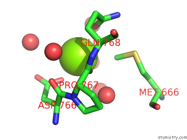
Mono view

Stereo pair view
Mono view
Stereo pair view
A full contact list of Magnesium with other atoms in the Mg binding
site number 1 of Crystal Structure of the C-Terminal Truncated Alpha-Kinase Domain of Myosin Heavy Chain Kinase within 5.0Å range:
|
Magnesium binding site 2 out of 2 in 3pdt
Go back to
Magnesium binding site 2 out
of 2 in the Crystal Structure of the C-Terminal Truncated Alpha-Kinase Domain of Myosin Heavy Chain Kinase
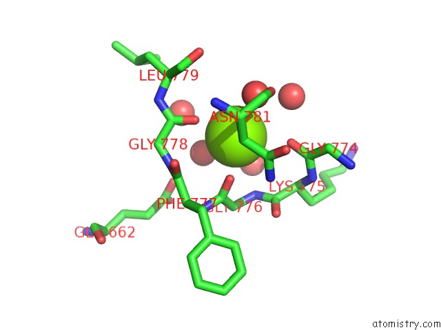
Mono view
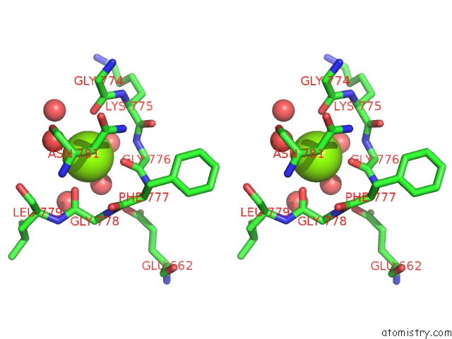
Stereo pair view
Mono view
Stereo pair view
A full contact list of Magnesium with other atoms in the Mg binding
site number 2 of Crystal Structure of the C-Terminal Truncated Alpha-Kinase Domain of Myosin Heavy Chain Kinase within 5.0Å range:
|
Reference:
S.W.Crawley,
M.S.Gharaei,
Q.Ye,
Y.Yang,
B.Raveh,
N.London,
O.Schueler-Furman,
Z.Jia,
G.P.Cote.
Autophosphorylation Activates Dictyostelium Myosin II Heavy Chain Kinase A By Providing A Ligand For An Allosteric Binding Site in the {Alpha}-Kinase Domain. J.Biol.Chem. V. 286 2607 2011.
ISSN: ISSN 0021-9258
PubMed: 21071445
DOI: 10.1074/JBC.M110.177014
Page generated: Thu Aug 15 09:11:04 2024
ISSN: ISSN 0021-9258
PubMed: 21071445
DOI: 10.1074/JBC.M110.177014
Last articles
Ca in 2Z2YCa in 2Z30
Ca in 2Z2Z
Ca in 2Z2X
Ca in 2Z0J
Ca in 2Z2D
Ca in 2YZ7
Ca in 2YN3
Ca in 2YZ4
Ca in 2YOC