Magnesium »
PDB 3p1g-3pio »
3pf5 »
Magnesium in PDB 3pf5: Crystal Structure of Bs-Cspb in Complex with RU6
Protein crystallography data
The structure of Crystal Structure of Bs-Cspb in Complex with RU6, PDB code: 3pf5
was solved by
R.Sachs,
K.E.A.Max,
U.Heinemann,
with X-Ray Crystallography technique. A brief refinement statistics is given in the table below:
| Resolution Low / High (Å) | 17.46 / 1.68 |
| Space group | P 21 21 21 |
| Cell size a, b, c (Å), α, β, γ (°) | 49.038, 49.769, 57.380, 90.00, 90.00, 90.00 |
| R / Rfree (%) | 18 / 23.5 |
Magnesium Binding Sites:
The binding sites of Magnesium atom in the Crystal Structure of Bs-Cspb in Complex with RU6
(pdb code 3pf5). This binding sites where shown within
5.0 Angstroms radius around Magnesium atom.
In total only one binding site of Magnesium was determined in the Crystal Structure of Bs-Cspb in Complex with RU6, PDB code: 3pf5:
In total only one binding site of Magnesium was determined in the Crystal Structure of Bs-Cspb in Complex with RU6, PDB code: 3pf5:
Magnesium binding site 1 out of 1 in 3pf5
Go back to
Magnesium binding site 1 out
of 1 in the Crystal Structure of Bs-Cspb in Complex with RU6
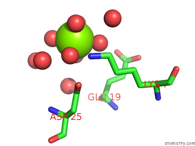
Mono view
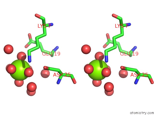
Stereo pair view
Mono view
Stereo pair view
A full contact list of Magnesium with other atoms in the Mg binding
site number 1 of Crystal Structure of Bs-Cspb in Complex with RU6 within 5.0Å range:
|
Reference:
R.Sachs,
K.E.Max,
U.Heinemann,
J.Balbach.
Rna Single Strands Bind to A Conserved Surface of the Major Cold Shock Protein in Crystals and Solution. Rna V. 18 65 2012.
ISSN: ISSN 1355-8382
PubMed: 22128343
DOI: 10.1261/RNA.02809212
Page generated: Thu Aug 15 09:13:26 2024
ISSN: ISSN 1355-8382
PubMed: 22128343
DOI: 10.1261/RNA.02809212
Last articles
Zn in 9MJ5Zn in 9HNW
Zn in 9G0L
Zn in 9FNE
Zn in 9DZN
Zn in 9E0I
Zn in 9D32
Zn in 9DAK
Zn in 8ZXC
Zn in 8ZUF